Jump to content
In this section we have spot diagnoses posted on a daily basis since June 2010, now over 4000! You can review the archived cases and read the suggested diagnoses by users and the final comment by the contributors.
Case are uploaded each week day by 10 am UK time with the correct diagnosis will generally be posted at 8 pm UK time. Why not view the most recent spot diagnosis and proffer a diagnosis?

Case Number : Case 564 - 7 Aug Posted By: Guest

Please read the clinical history and view the images by clicking on them before you proffer your diagnosis.
Submitted Date :
   (0 reviews)

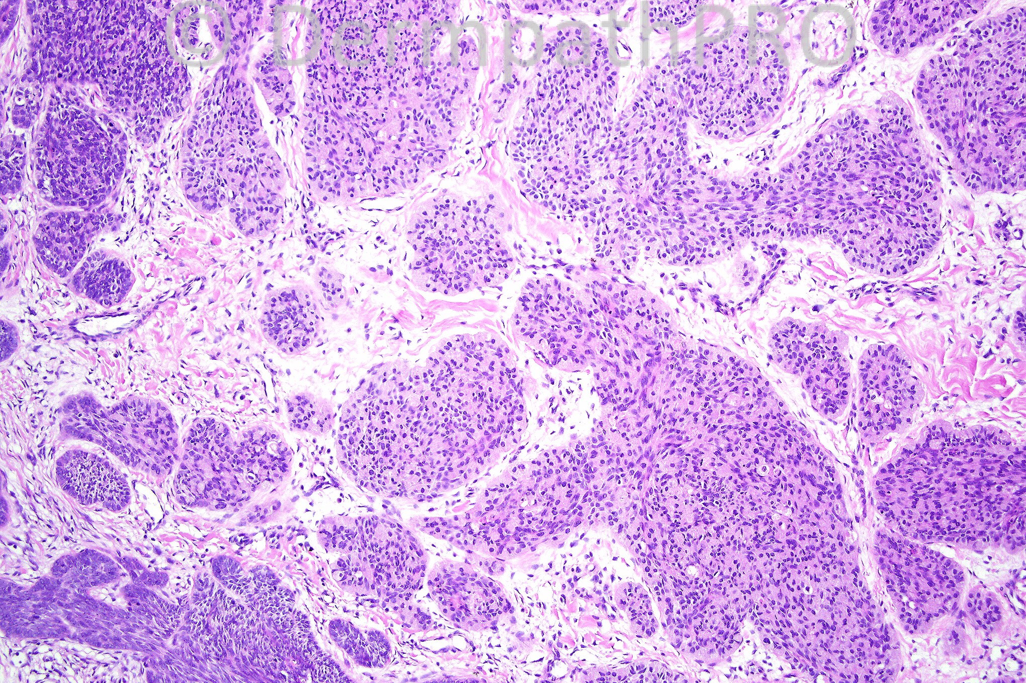
Female 82 years with a 2.0 cm nodule on head.

We are grateful to Dr. Wayne Grayson who has provided this case.


  Report Record

User Feedback


Guest Hazem Hamed

Posted

Metastatic poorly differentiated adenocarcinoma (likely from the breast). Clinicoradiological correlation and immuno to identify the primary. If no primary could be identified I would consider porocarcinoma.

Share this comment


Link to comment
share_externally

Dr. Mona Abdel-Halim

Posted

I think this is a very interesting case. Careful examination of images 1 & 2 will show some islands with peripheral palisdaing. The 3rd image however shows clearly that the cells have esinophilic granular cytoplasm. I will diagnose this as granular cell basal cell carcinoma in which according to McKee's Pathology of the skin, some if not all the neoplastic cells may have abundant granular esinophilic cytoplasm due to membrane bound lysosome like granules.

Share this comment


Link to comment
share_externally

Guest Dr. Francisco Vílchez

Posted

[b]Basal cell carcinoma. [/b]Agree with Mona, there is clearly peripheral palisading, especially in pic. 2.

Share this comment


Link to comment
share_externally

Guest Dr Gonzalo de Toro

Posted

Granular basal cell carcinoma

Share this comment


Link to comment
share_externally

Guest Hazem Hamed

Posted

I aggree with Granular basal cell carcinoma as DD. I can see suggestions of intracytoplasmic lumina which I think are not true lumina but in fact are spaces left behind apoptotic cells.

Share this comment


Link to comment
share_externally

Guest MarieMD

Posted

BCC, granular cell variant. (periperhal islands are more classic BCC)

Share this comment


Link to comment
share_externally

Dr. Phillip McKee

Posted

Well done all of you who diagnosed granular cell BCC. I could have shown you a scanning view which would have made the diagnosis easier but I wanted to see how you would do with only the higher power views. Excellent.

Share this comment


Link to comment
share_externally

Guest Sadiq M Amer

Posted

[color=#1C2837][font=arial, verdana, tahoma, sans-serif][size=4]I am in favor of granular basal cell carcinoma, the differential include [/size][/font][/color]cellular neurothekeoma

and granular cell tumor.

Share this comment


Link to comment
share_externally


×
×
  • Create New...