Jump to content
In this section we have spot diagnoses posted on a daily basis since June 2010, now over 4000! You can review the archived cases and read the suggested diagnoses by users and the final comment by the contributors.
Case are uploaded each week day by 10 am UK time with the correct diagnosis will generally be posted at 8 pm UK time. Why not view the most recent spot diagnosis and proffer a diagnosis?

Case Number : Case 565 - 8 Aug Posted By: Guest

Please read the clinical history and view the images by clicking on them before you proffer your diagnosis.
Submitted Date :
   (0 reviews)

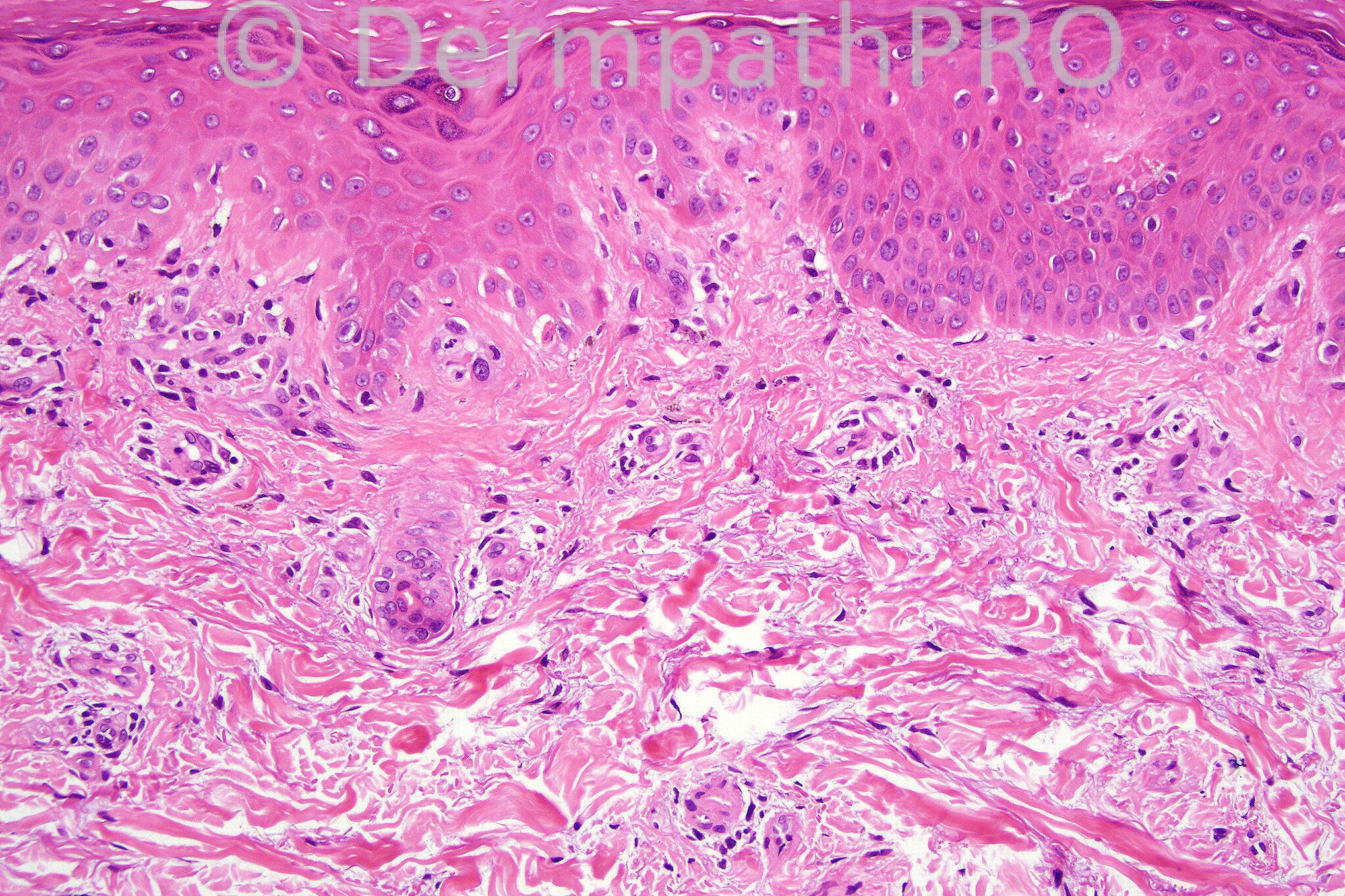
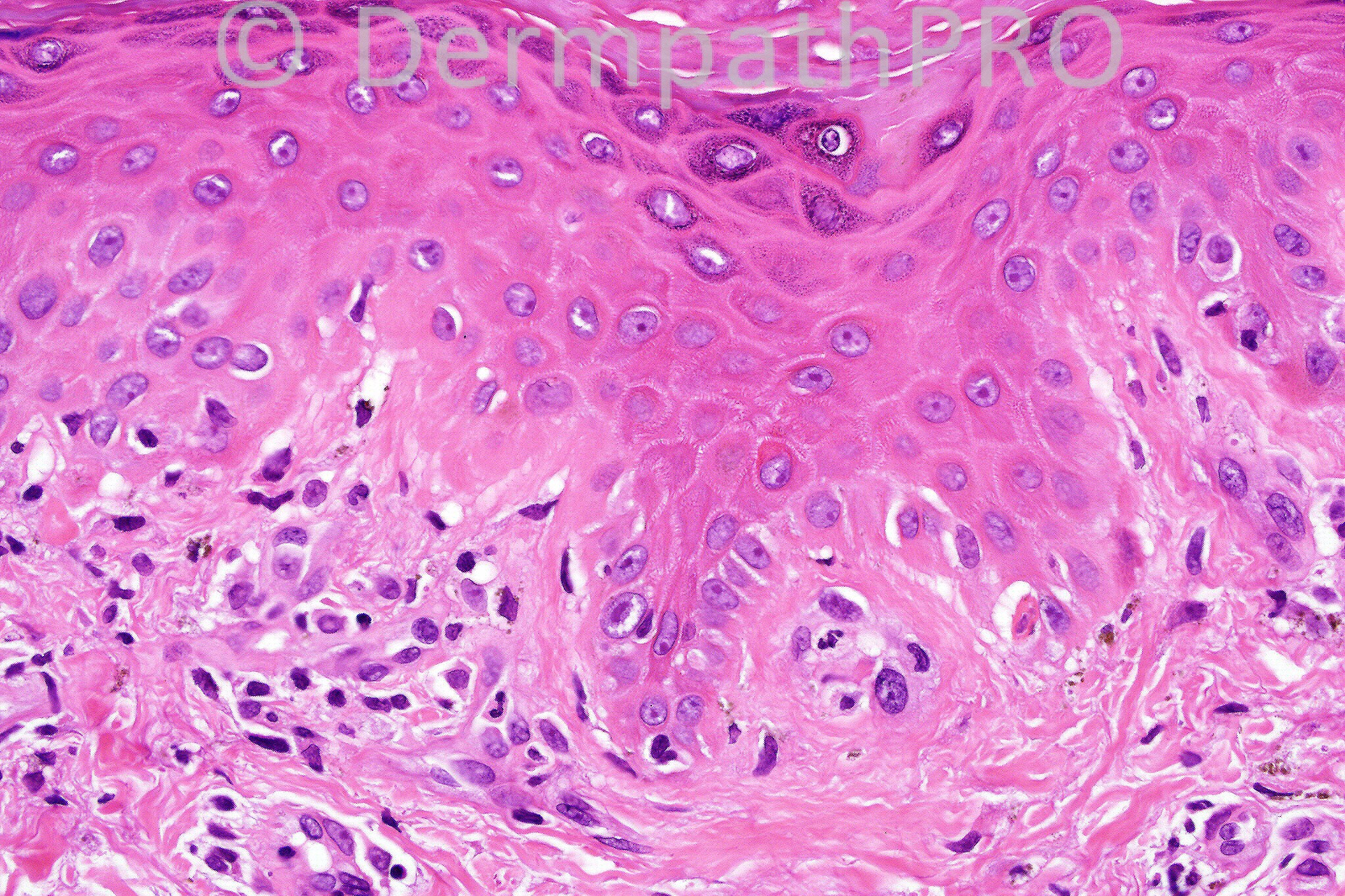
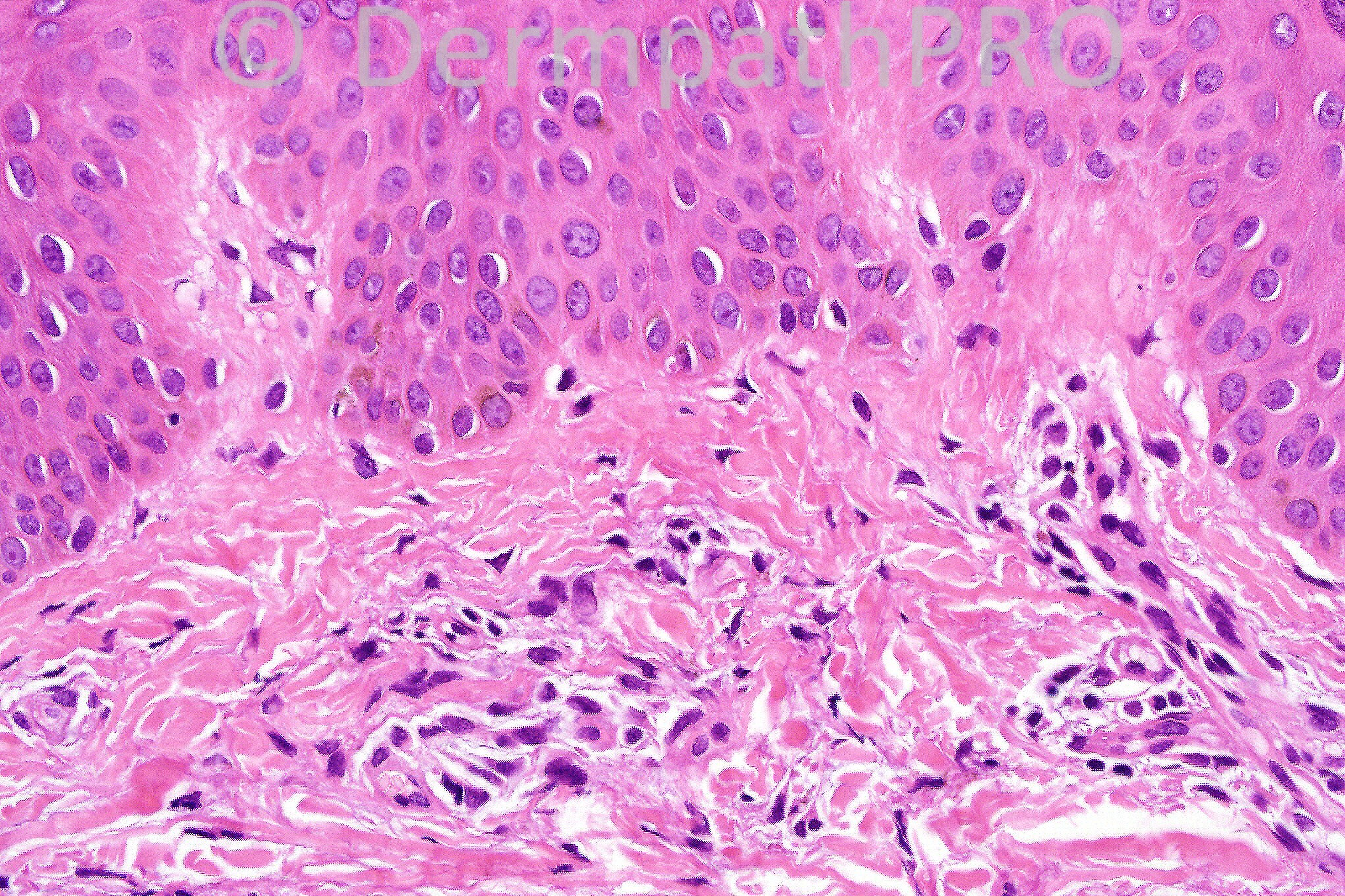
Periorbital swelling.  Erythematous rash forearms, knuckle of fingers.

We are grateful to Dr. Richard Carr who has provided this case.


  Report Record

User Feedback


Guest Hazem Hamed

Posted

Mild vacuolar interface reaction pattern, with the history provided, would be consistent with dermatomyositis. Correlation with serology for anti Jo-1 and Ro/SS-A antibodies is required. Also the patient should be investigated for possible underlying malignancy.

Share this comment


Link to comment
share_externally

Guest Dr. Francisco Vílchez

Posted

Dermatomyositis.

Share this comment


Link to comment
share_externally

Sasi Attili

Posted

dermatomyositis

Share this comment


Link to comment
share_externally

Dr. Mona Abdel-Halim

Posted

Dermatomyositis.

Share this comment


Link to comment
share_externally

Guest Dr Gonzalo de Toro

Posted

Dermatomyositis. The most consistent histological findings of DM included increased dermal mucin, vacuolar alteration of the basal cell layer, and mild-to-moderate mononuclear cell inflammatory infiltrates.

Share this comment


Link to comment
share_externally

Guest MarieMD

Posted

changes of dermatomyositis, given clinical

Share this comment


Link to comment
share_externally

Dr. Phillip McKee

Posted

Although the differential diagnosis of interface change is wide, given the clinical information, I agree with the diagnosis of dermatomyositis. Although I do not have the full clinical information and in particular the site of the specimen, the stratum corneum in fig 1 suggests a distal acral site and I think that we might be looking ar a Gottron's papule

Share this comment


Link to comment
share_externally


×
×
  • Create New...