Jump to content
In this section we have spot diagnoses posted on a daily basis since June 2010, now over 4000! You can review the archived cases and read the suggested diagnoses by users and the final comment by the contributors.
Case are uploaded each week day by 10 am UK time with the correct diagnosis will generally be posted at 8 pm UK time. Why not view the most recent spot diagnosis and proffer a diagnosis?

Case Number : Case 567 - 10 Aug Posted By: Guest

Please read the clinical history and view the images by clicking on them before you proffer your diagnosis.
Submitted Date :
   (0 reviews)

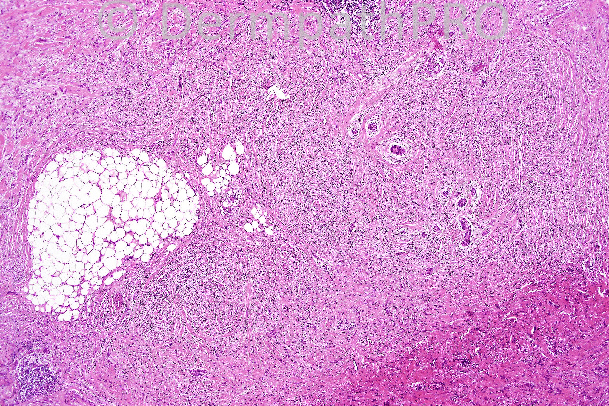
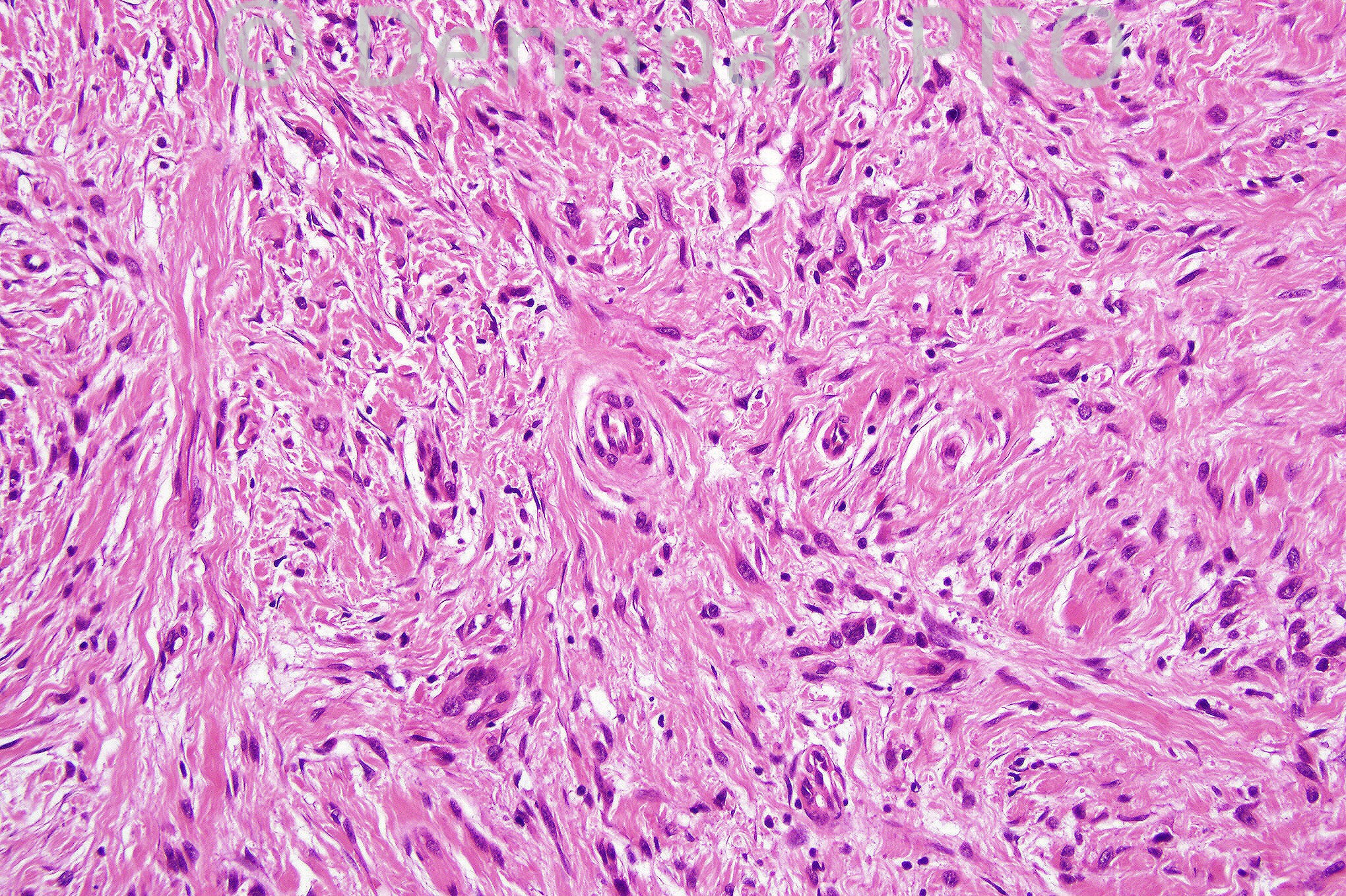
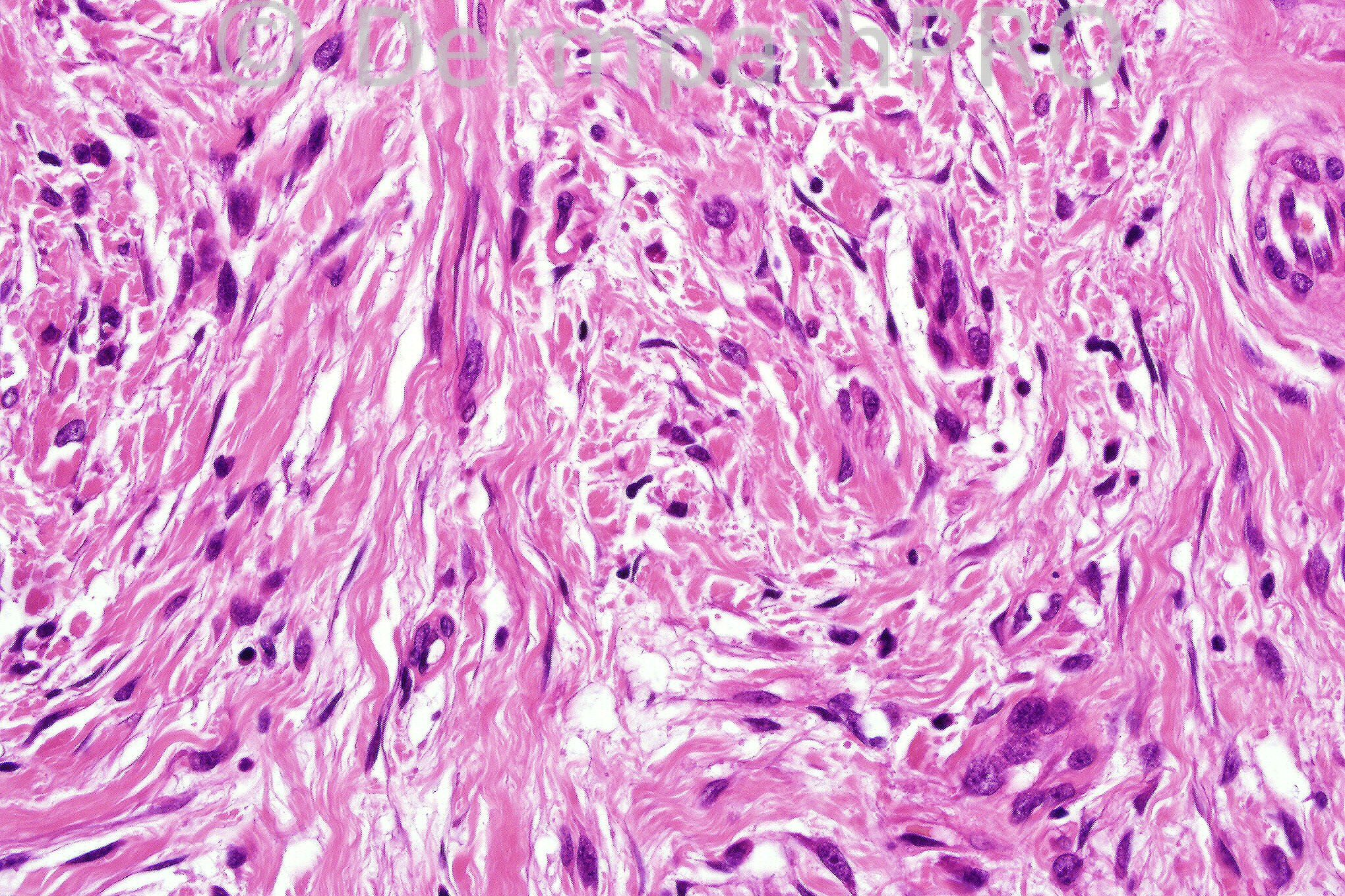
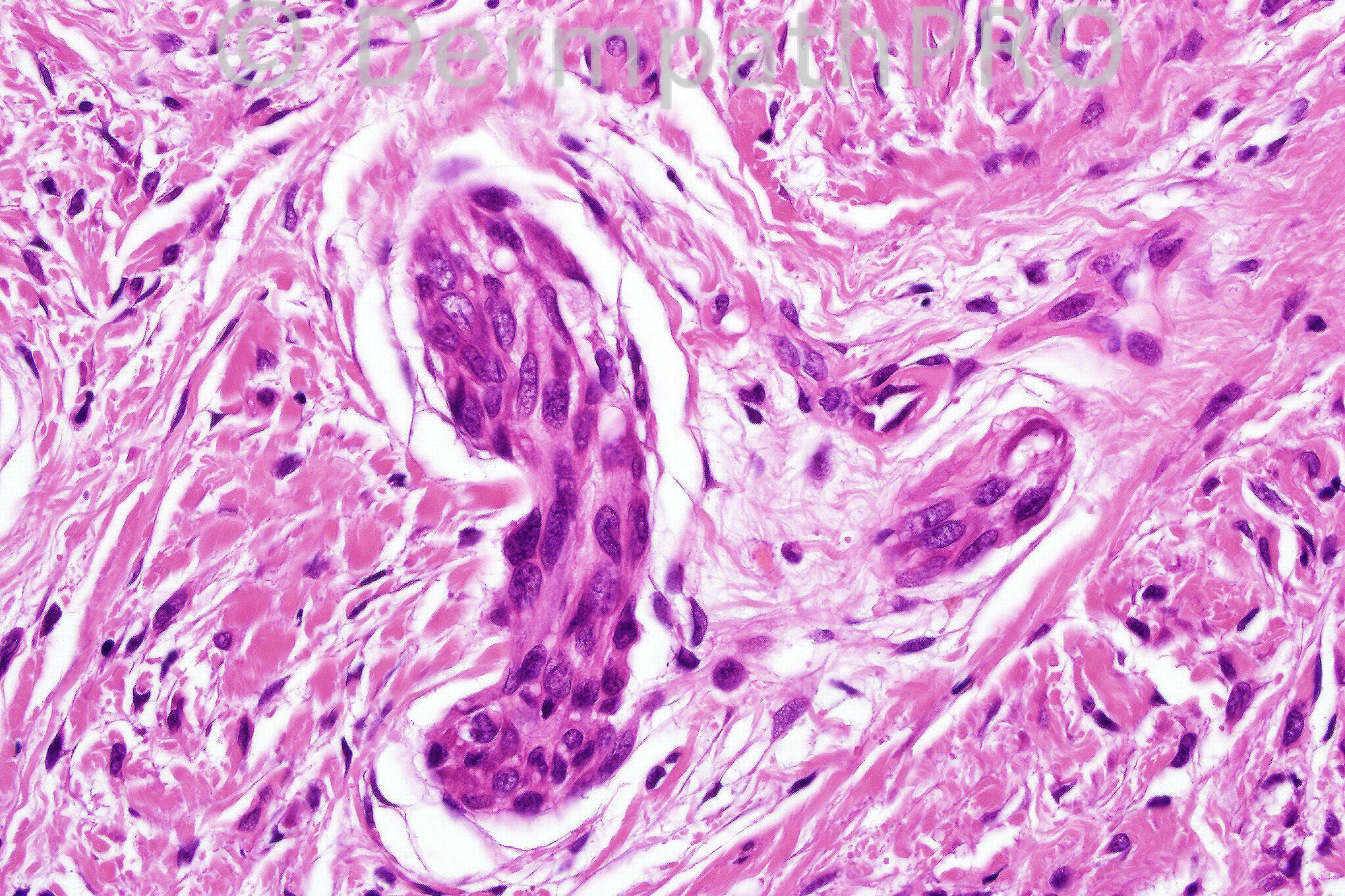
Female 72 years with a lesion on forehead.


  Report Record

User Feedback


Guest Hazem Hamed

Posted

Difficult case but I would give a differential: Desmoplastic melanoma, Dermatofibrosarcoma protuberance, extra-acral perineurioma and angiofibroma for immunohistochemistry.

Share this comment


Link to comment
share_externally

Dr. Mona Abdel-Halim

Posted

Agree with Dr Hazem that this is a difficult case. I thought of desmoplastic melanoma. I guess the lymphocytic collections in Fig 1 represent a clue. I still need to confirm with immunos.

Share this comment


Link to comment
share_externally

Guest MarieMD

Posted

Though @ first desmoplastic melanoma comes to mind (low power), the higher power views, especially foto 4, have features of leiomyosarcoma. I would still do immunos.

Share this comment


Link to comment
share_externally

Guest Eman El-Nabarawy

Posted

Thought of spindle cell AFX or extra-acral perineuroma for immunos.

Share this comment


Link to comment
share_externally

spindly malignant tumor for immuno !!! MPNST? in Fig. 4 is this metastasis ?? seems as if it is within a vessel.

Share this comment


Link to comment
share_externally

Guest A Bansal

Posted

DD: DFSP, MM, Spindle cell carcinoma, AFX. Immuno needed. If image 4 shows an entrapped adnexal structure, does that alone make any of the above such as AFX more likely?

Share this comment


Link to comment
share_externally

Dr. Phillip McKee

Posted

Thank you for all of your suggestions. I believe that desmoplastic melanoma is the correct diagnosis. The nested population in the 4th figure is very suggestive of a melanocytic population and the lymphoid aggregate could be a diagnostic clue. This case is an old one when immunohistochemistry hadn't even been though of!! After some effort, I did discover that the patient had had a desmolastic melanoma excised previously at that site. Have a great weekend and many thanks for your continued support.

Share this comment


Link to comment
share_externally


×
×
  • Create New...