Jump to content
In this section we have spot diagnoses posted on a daily basis since June 2010, now over 4000! You can review the archived cases and read the suggested diagnoses by users and the final comment by the contributors.
Case are uploaded each week day by 10 am UK time with the correct diagnosis will generally be posted at 8 pm UK time. Why not view the most recent spot diagnosis and proffer a diagnosis?

Case Number : Case 578 - 27 Aug Posted By: Guest

Please read the clinical history and view the images by clicking on them before you proffer your diagnosis.
Submitted Date :
   (0 reviews)

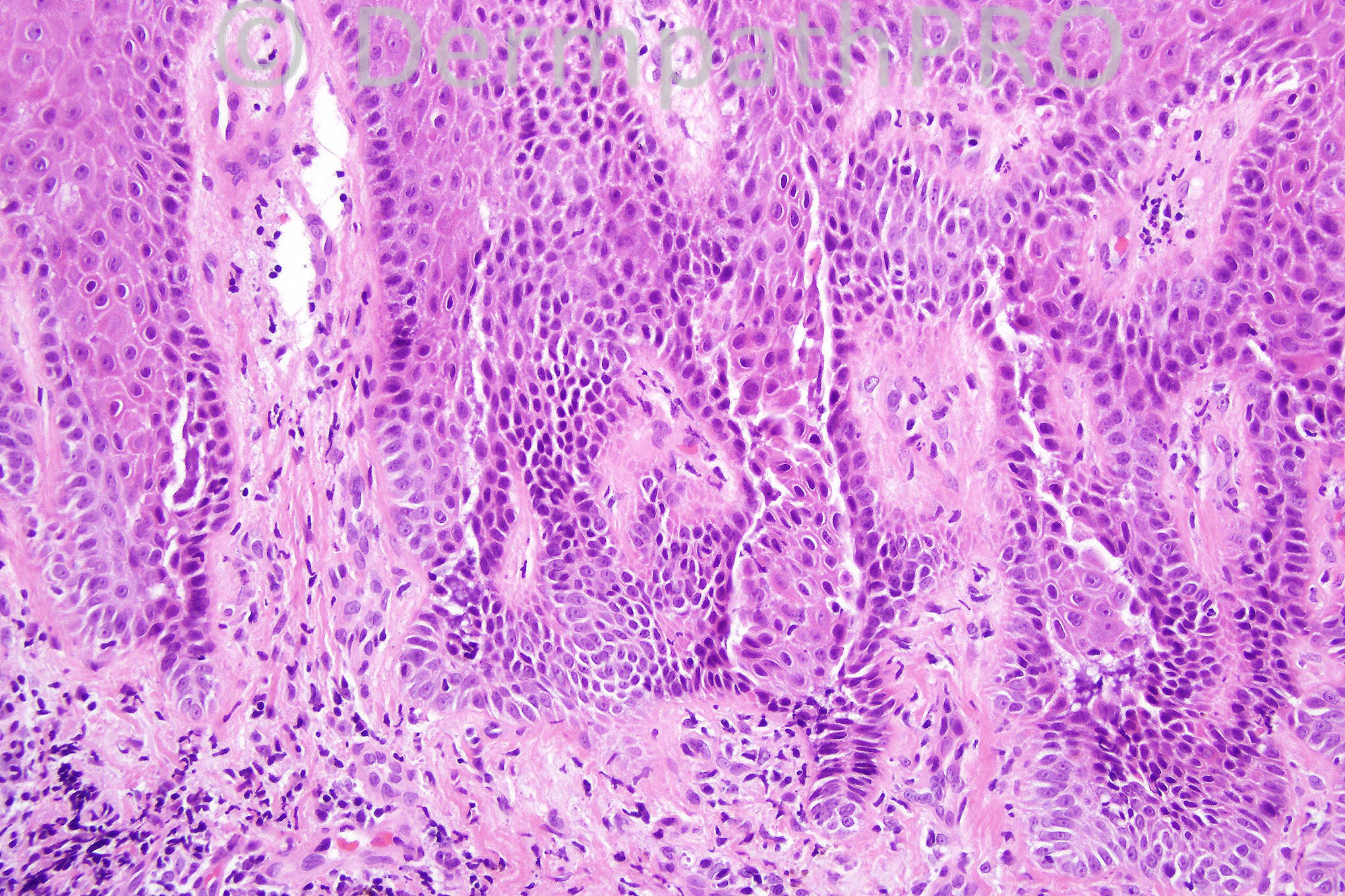
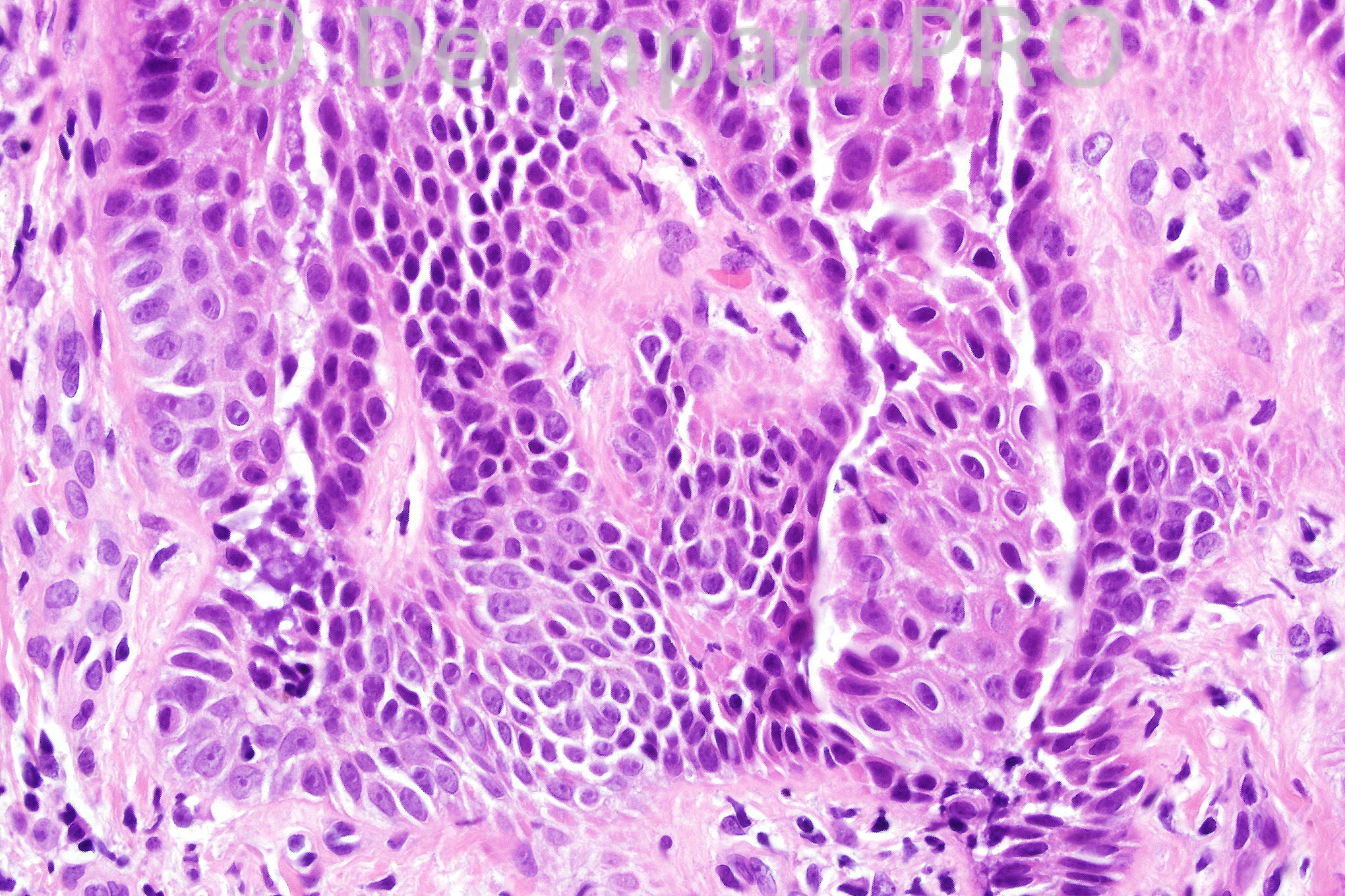
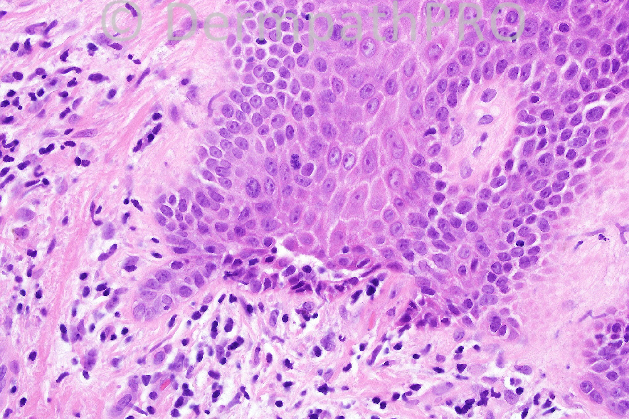
Male 67 years with blisters on the trunk.


  Report Record

User Feedback


Guest Dr. Francisco Vílchez

Posted

Suprabasal clefting. No eosinophils. Pemphigus vs. Grover´s.

Share this comment


Link to comment
share_externally

Dr. Mona Abdel-Halim

Posted

[b]Focal Suprabasal clefting, no esinophils, pathologically favors Grover's. Still on clinical basis, pemphigus is favored. [/b]

Share this comment


Link to comment
share_externally

Sasi Attili

Posted

Looks like Grover's. But the clinical history of blisters is unusual for Grover's. Need DIF studies to differentiate from pemphigus therefore.

Share this comment


Link to comment
share_externally

Guest MarieMD

Posted

focal acantholytic dyskeratosis c/w Grover's dz in this setting.

Share this comment


Link to comment
share_externally

Guest Dr Engin Sezer

Posted

I feel that small size of the lesion (involvement of just a few rete ridges) may be a clue for pemphigus-like variant of Grover's disease to differentiate from P. vulgaris. I'm curious about the Immuno results though.

Share this comment


Link to comment
share_externally

Dr. King-Chung Lee

Posted

In the low power picture, there are zones of pale suggesting spongiosis. I favor Grover's disease. Blisters, though rare, is possible in Grover.

Share this comment


Link to comment
share_externally

Guest Susan YH Shih

Posted

I see acantholysis with dyskeratosis, although no typical corps ronds or grains found. Besides, focal tombstone appearance is noted. Based on multiple patterns found in a single lesion, I wound say Grover's disease. A DIF is crucial still.

Share this comment


Link to comment
share_externally

Iskander H. Chaudhry

Posted

The diagnosis is paraneoplastic pemphigus. All the differentials are acceptable, the final diagnosis was based upon immunofluorescence and correlation with the clinical history.

Share this comment


Link to comment
share_externally


×
×
  • Create New...