Jump to content
In this section we have spot diagnoses posted on a daily basis since June 2010, now over 4000! You can review the archived cases and read the suggested diagnoses by users and the final comment by the contributors.
Case are uploaded each week day by 10 am UK time with the correct diagnosis will generally be posted at 8 pm UK time. Why not view the most recent spot diagnosis and proffer a diagnosis?

Case Number : Case 648 - 3 Dec Posted By: Guest

Please read the clinical history and view the images by clicking on them before you proffer your diagnosis.
Submitted Date :
   (0 reviews)

None. Case included for interest, courtesy of Dr. Thomas Brenn.


  Report Record

User Feedback


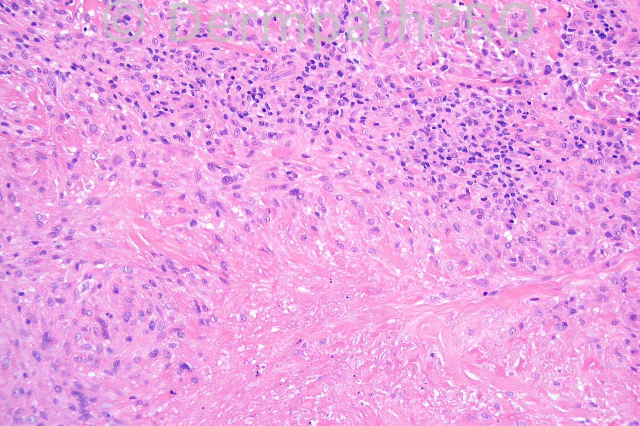
Palisaded granulomatous dermatitis with necrobiosis mucin deposition and some fibrinoid necrosis: granuloma annulare vs rheumatoid nodule

Share this comment


Link to comment
share_externally

Sasi Attili

Posted

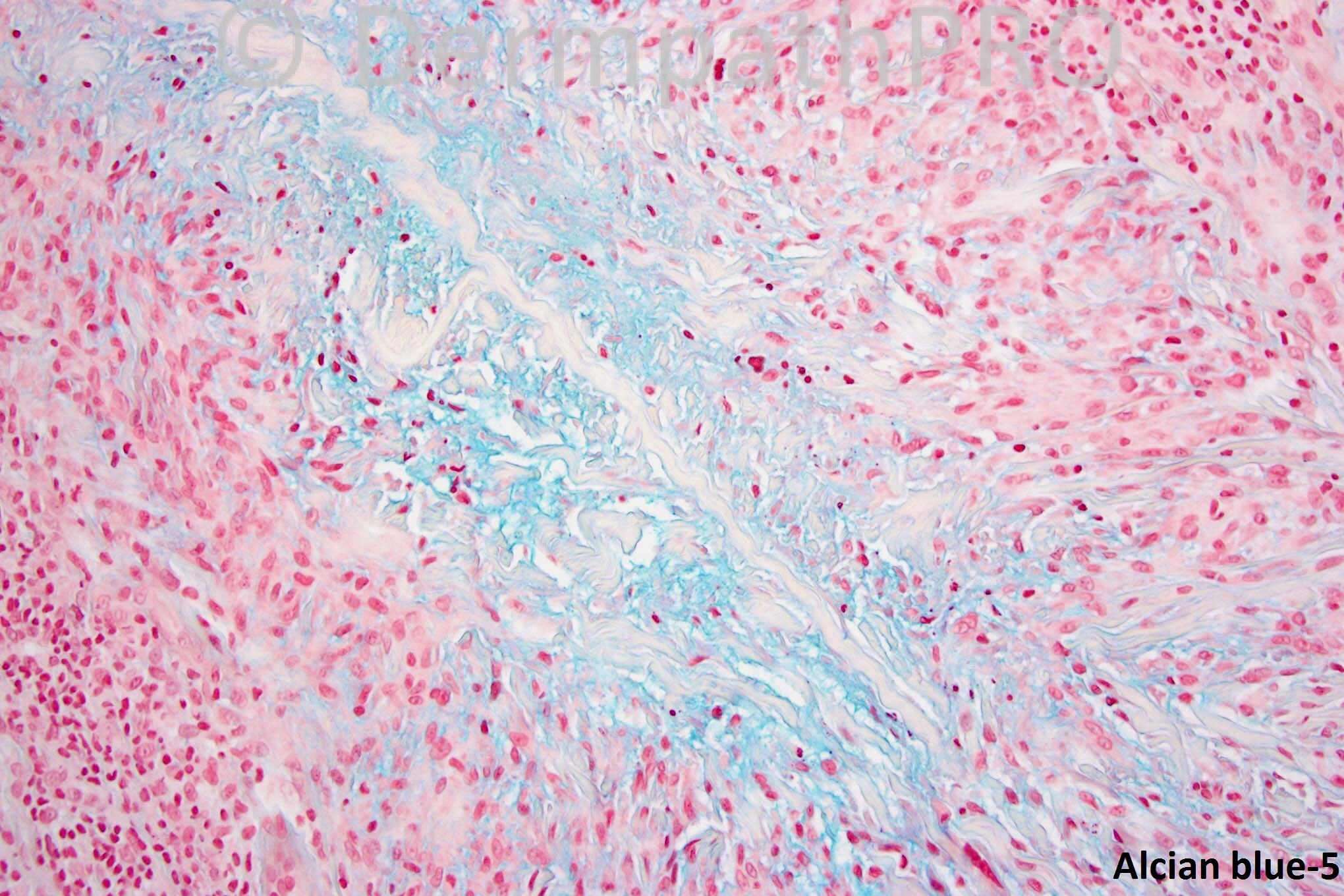
Deep GA favoured over RN, due to mucin ++.

Share this comment


Link to comment
share_externally

Guest Dr Engin Sezer

Posted

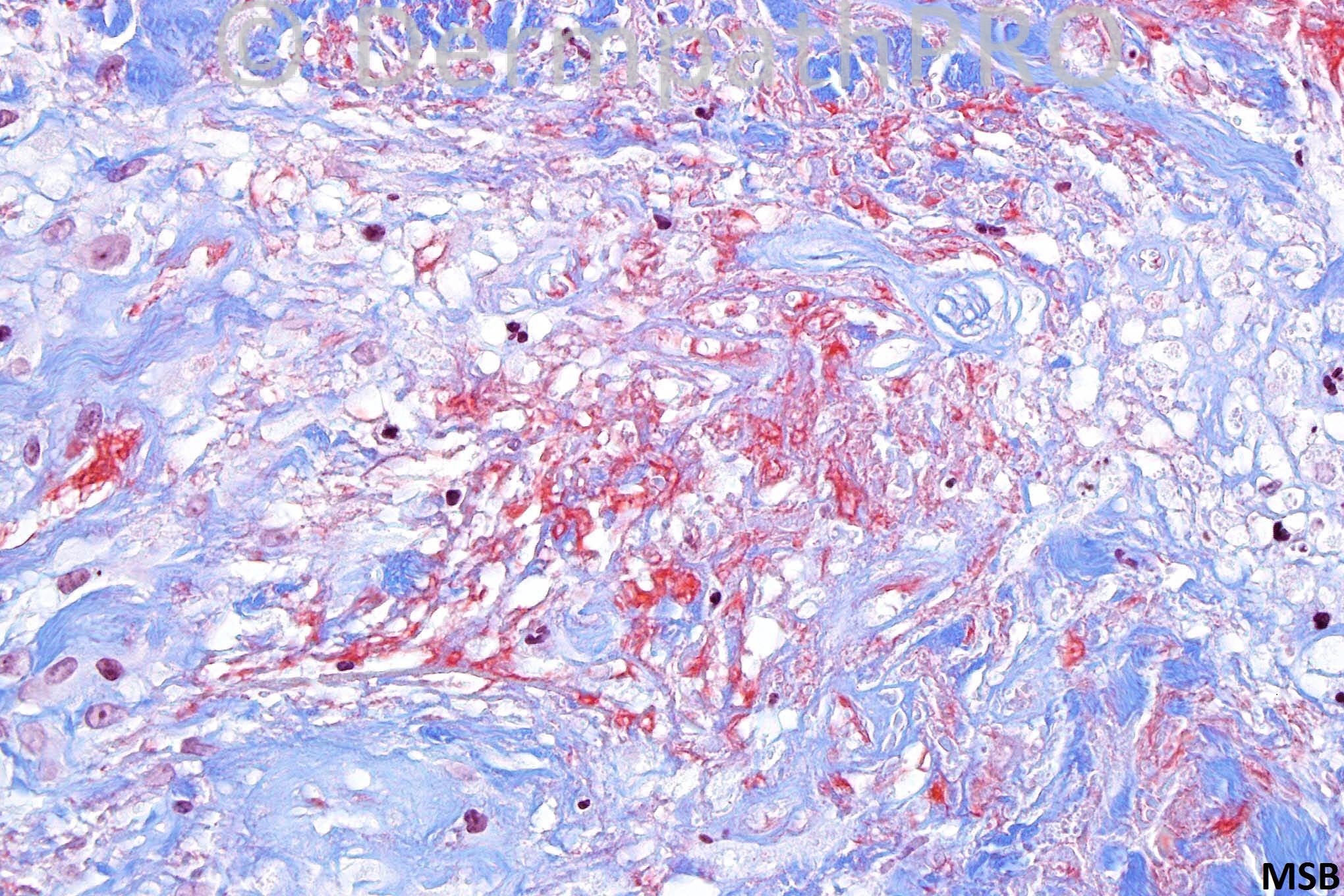
I agree with Dr. Saeb with DDX including GA vs. rheumatoid nodule. I favor GA over rheumatoid nodule,since the cases that I've seen with latter diagnosis showed more prominent fibrin in H&E sections, however lack of history of a rheumatological disorder may be helpful to entirely rule out the diagnosis.

Share this comment


Link to comment
share_externally

I signed out two cases this past week with just this same differential, both deep-seated necrobiotic granulomatous processes with + mucin. One was a 22yo female with no rheumatoid arthritis history, the other a 58yo male again with no RA history. The 58yo male also had what appeared to be classic GA w/ some interstitial features in the overlying dermis. I favored subQ GA for both with a differential of rheumatoid nodule.

Share this comment


Link to comment
share_externally

Dr. Phillip McKee

Posted

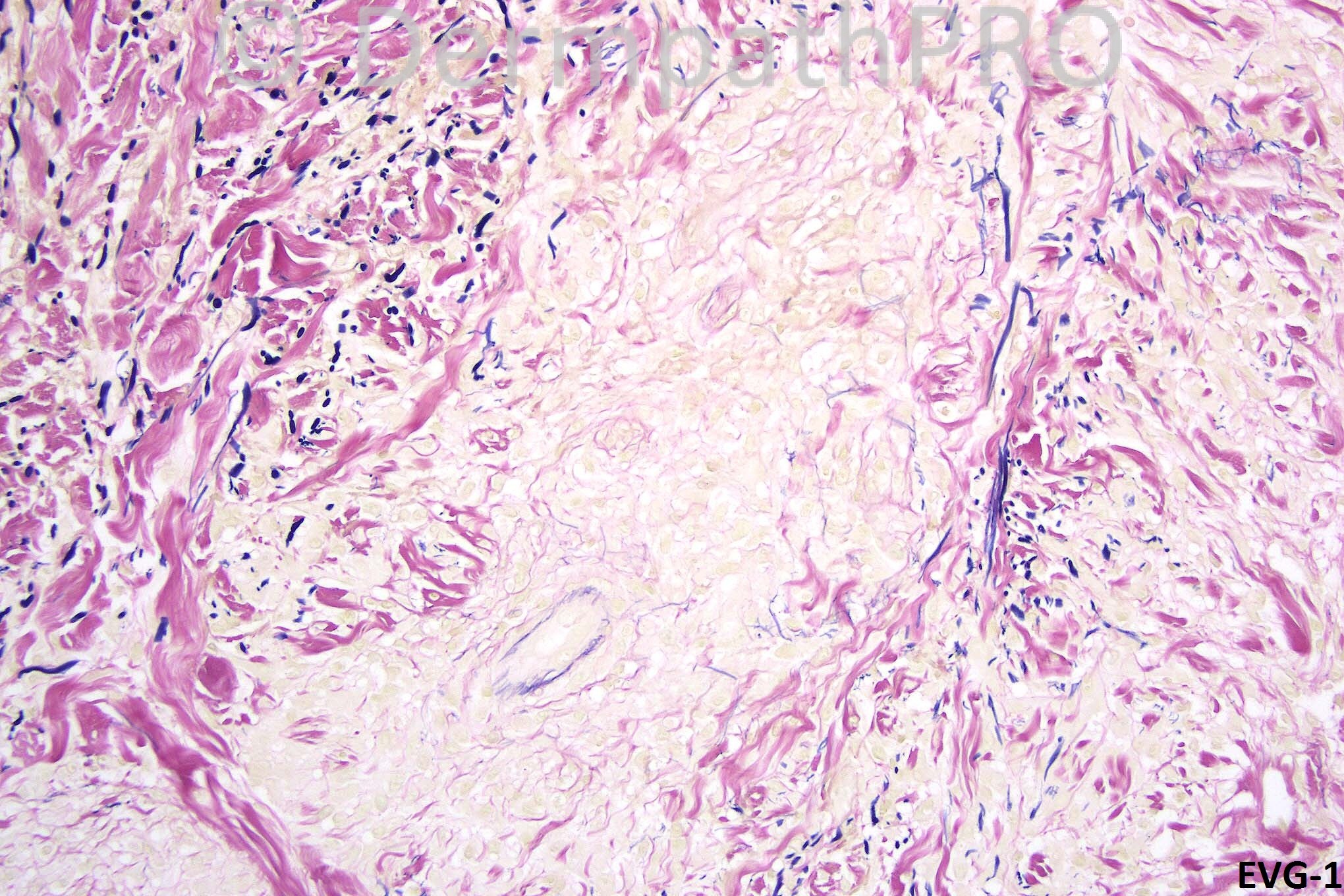
I included this case to show the fibrin, mucin and elastic tissue stains in GA. It is not often that one sees such beautiful examples. Many thanks Thomas.

Share this comment


Link to comment
share_externally

Guest Robledo F. Rocha

Posted

A large area of necrobiosis is surrounded by a palisade of lymphocytes and histiocytes. The central necrobiotic area contain increased amounts of mucin that characterizes it as a “blue granuloma” in contrast to the “red granuloma” with central fibrinoid necrosis. Also, there are no multinucleated histiocytes within the palisaded granuloma. So, I think it’s a granuloma annulare.

Share this comment


Link to comment
share_externally

Guest MarieMD

Posted

GA, great example with nice mucin staining

Share this comment


Link to comment
share_externally


×
×
  • Create New...