Jump to content
In this section we have spot diagnoses posted on a daily basis since June 2010, now over 4000! You can review the archived cases and read the suggested diagnoses by users and the final comment by the contributors.
Case are uploaded each week day by 10 am UK time with the correct diagnosis will generally be posted at 8 pm UK time. Why not view the most recent spot diagnosis and proffer a diagnosis?

Case Number : Case 650 - 5 Dec Posted By: Guest

Please read the clinical history and view the images by clicking on them before you proffer your diagnosis.
Submitted Date :
   (0 reviews)

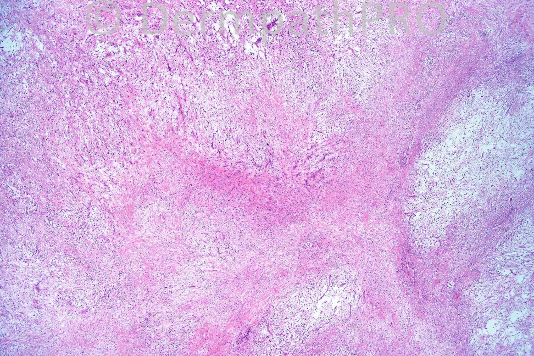
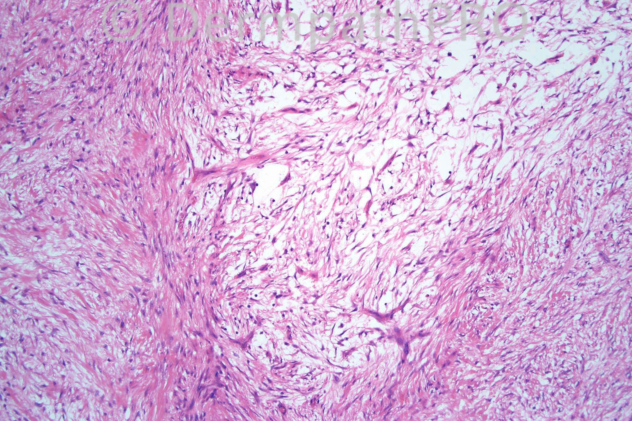
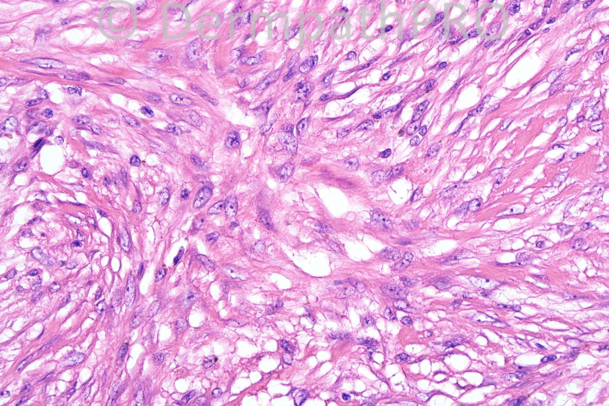
Female 27 years with a 1.0 cm nodule on the forearm.


  Report Record

User Feedback


Guest Dr Engin Sezer

Posted

Agree nodular fasciitis

Share this comment


Link to comment
share_externally

but RBCs xtravasation, lymphocytic infiltrate are not obvious??

Share this comment


Link to comment
share_externally

Sasi Attili

Posted

Nodular fasciitis sine extravasated RBC!

Share this comment


Link to comment
share_externally

Dr. Mona Abdel-Halim

Posted

First impression was nodular fasciitis due to the myxoid areas with feathering but could not find any extravasated erythrocytes.. Could be something else? Like low grade fibromyxoid sarcoma??

Share this comment


Link to comment
share_externally

Guest Robledo F. Rocha

Posted

Agree with all: nodular fasciitis. Swirling bundles of spindle-shaped cells set in a myxoid edematous background with microcystic degeneration and tissue cleavages.

Share this comment


Link to comment
share_externally

Guest Robledo F. Rocha

Posted

Agree with all: nodular fasciitis. Swirling bundles of spindle-shaped cells set in a myxoid edematous background with microcystic degeneration and tissue cleavages.

Share this comment


Link to comment
share_externally

Arif Usmani

Posted

Nodular fasciitis however agree with Mona's differential.

Share this comment


Link to comment
share_externally

Dr. Phillip McKee

Posted

Nodular fasciitis is correct. With respect to the red cell extravasation it probably was present but as I only take 4 images per case, I didn't photograph it. Also, it is important to remember that although any entity has characteristic features sometimes not all are present but this does not stop you making the diagnosis.

Share this comment


Link to comment
share_externally


×
×
  • Create New...