Jump to content
In this section we have spot diagnoses posted on a daily basis since June 2010, now over 4000! You can review the archived cases and read the suggested diagnoses by users and the final comment by the contributors.
Case are uploaded each week day by 10 am UK time with the correct diagnosis will generally be posted at 8 pm UK time. Why not view the most recent spot diagnosis and proffer a diagnosis?

Case Number : Case 652 - 7 Dec Posted By: Guest

Please read the clinical history and view the images by clicking on them before you proffer your diagnosis.
Submitted Date :
   (0 reviews)

None, case courtesy of Dr. Thomas Brenn


  Report Record

User Feedback


Guest Hazem Hamed

Posted

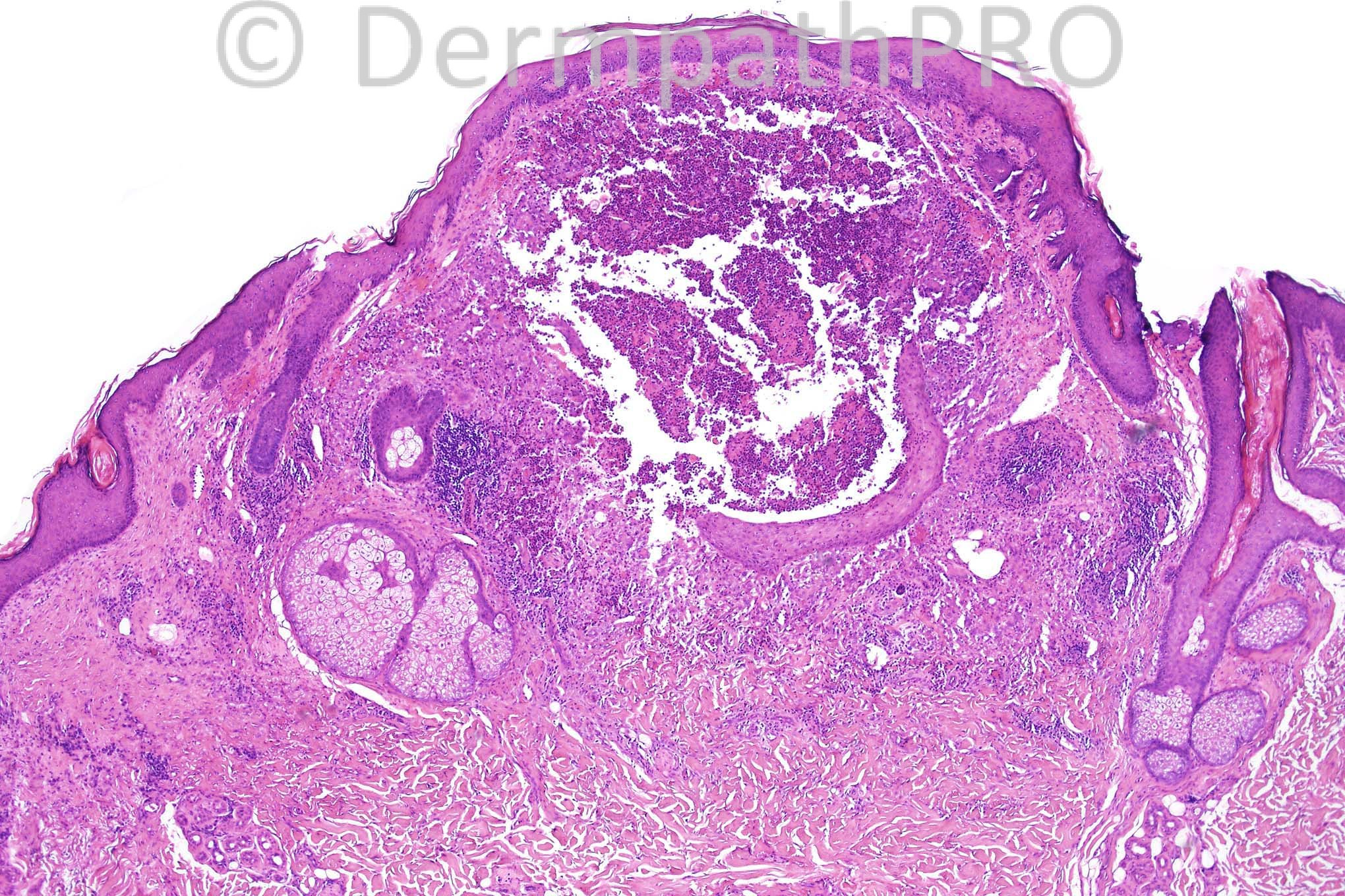
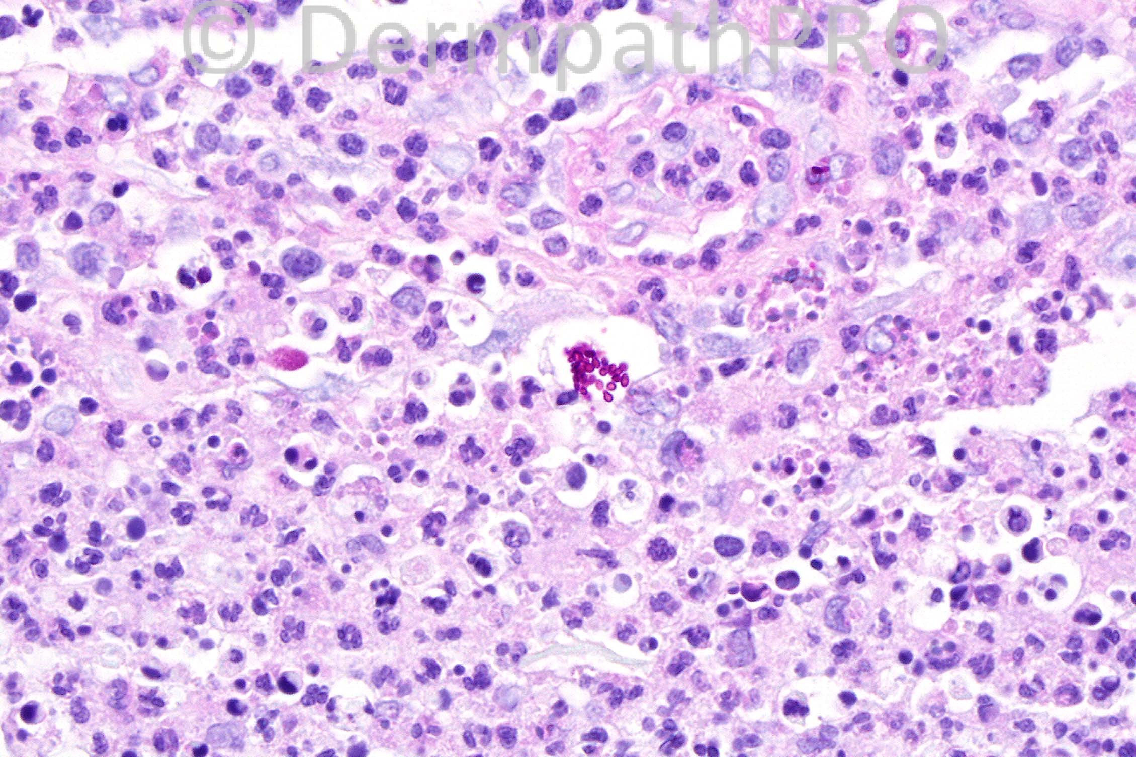
A suppurative granulomatous inflammation due to fungal infection. I think the spores are present within a spherule and not intracellular. So I would suggest Coccidioidomycosis for culture.

Share this comment


Link to comment
share_externally

Sasi Attili

Posted

[quote name='Hazem Hamed' timestamp='1354879854']
A suppurative granulomatous inflammation due to fungal infection. I think the spores are present within a spherule and not intracellular. So I would suggest Coccidioidomycosis for culture.
[/quote]
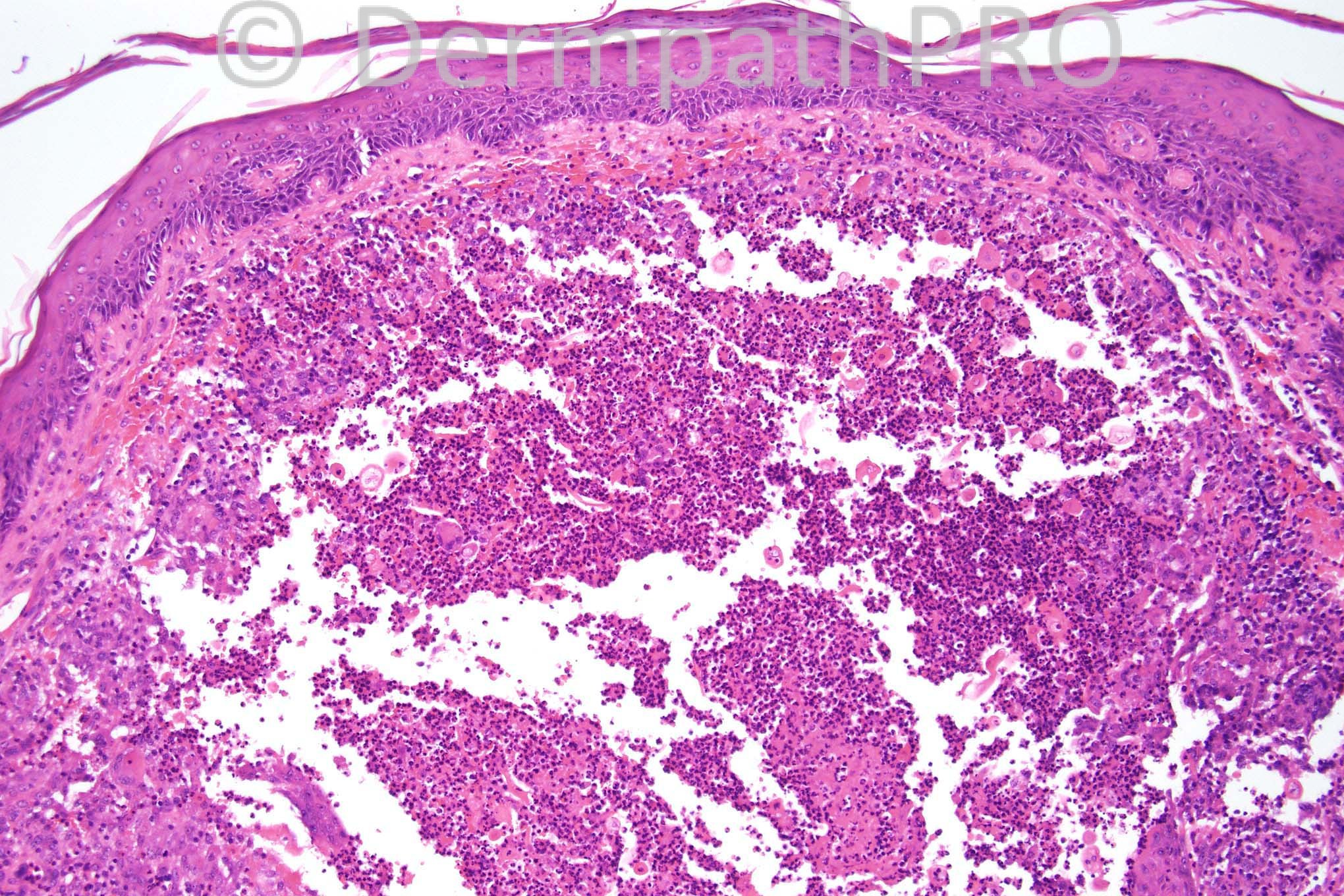
Agree that it is a suppurative granuloma, but not convinced that there is a spherule. To me it appears that the spores are just grouped together. Cultures are necessary. Are the spores probably a bit too big for pityrosporum?, which is a possibility.

Share this comment


Link to comment
share_externally

Guest Robledo F. Rocha

Posted

[font="Swiss721BT-Bold, sans-serif"][size="2"][size=1][font="Palatino Linotype, serif"][size="3"]Suppurative folliculitis with rupture due to a dermatophyte infection.[/size][/font][/size][/size][/font]

Share this comment


Link to comment
share_externally

Arif Usmani

Posted

I like the idea of pityrosporum folliculitis

Share this comment


Link to comment
share_externally

Dr. Mona Abdel-Halim

Posted

fungal folliculitis

Share this comment


Link to comment
share_externally

Dr. Phillip McKee

Posted

Well done Arif. The diagnosis is ptyrosporum folliculitis. The low power view shows residual distorted follicular epithelium. It may well be that the original section or deeper levels might have demonstrated the follicle to better effect. Thanks for your comments this week and have a wonderful weekend.

Share this comment


Link to comment
share_externally


×
×
  • Create New...