Jump to content
In this section we have spot diagnoses posted on a daily basis since June 2010, now over 4000! You can review the archived cases and read the suggested diagnoses by users and the final comment by the contributors.
Case are uploaded each week day by 10 am UK time with the correct diagnosis will generally be posted at 8 pm UK time. Why not view the most recent spot diagnosis and proffer a diagnosis?

Case Number : Case 654 - 11 Dec Posted By: Guest

Please read the clinical history and view the images by clicking on them before you proffer your diagnosis.
Submitted Date :
   (0 reviews)

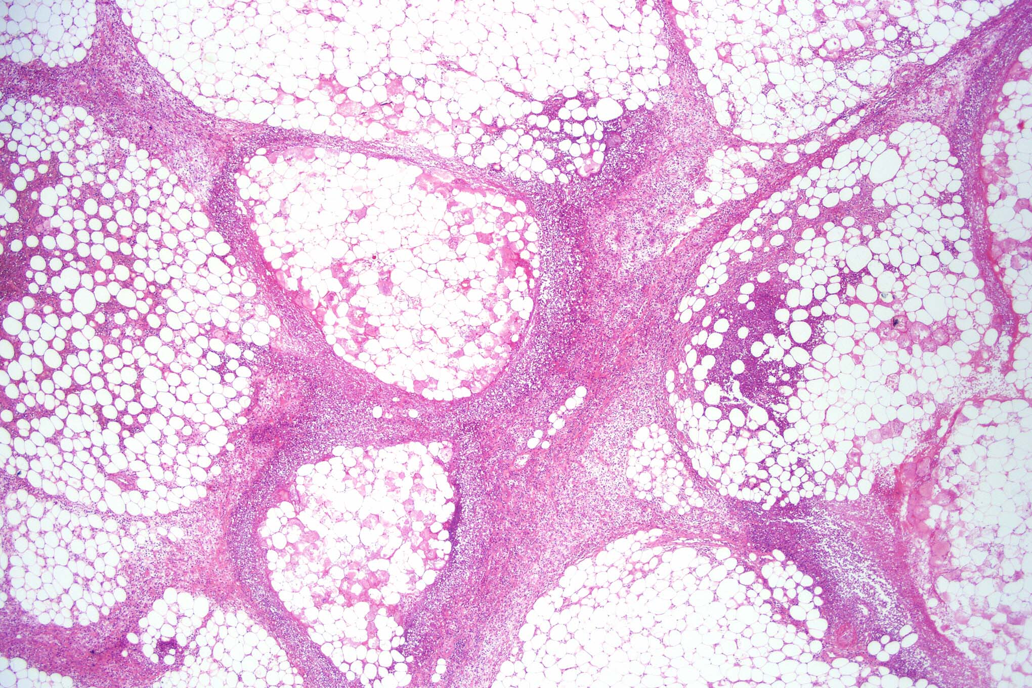
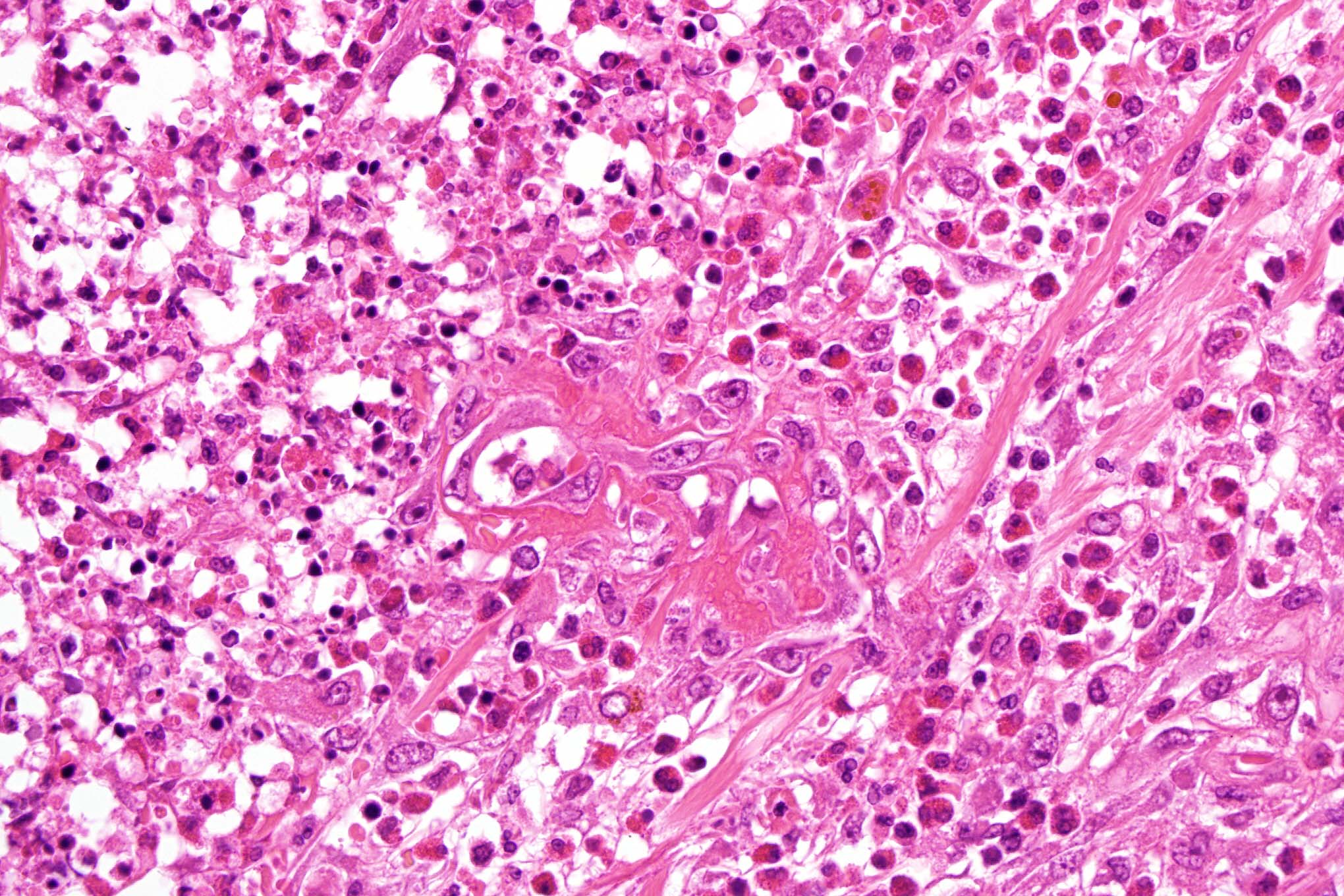
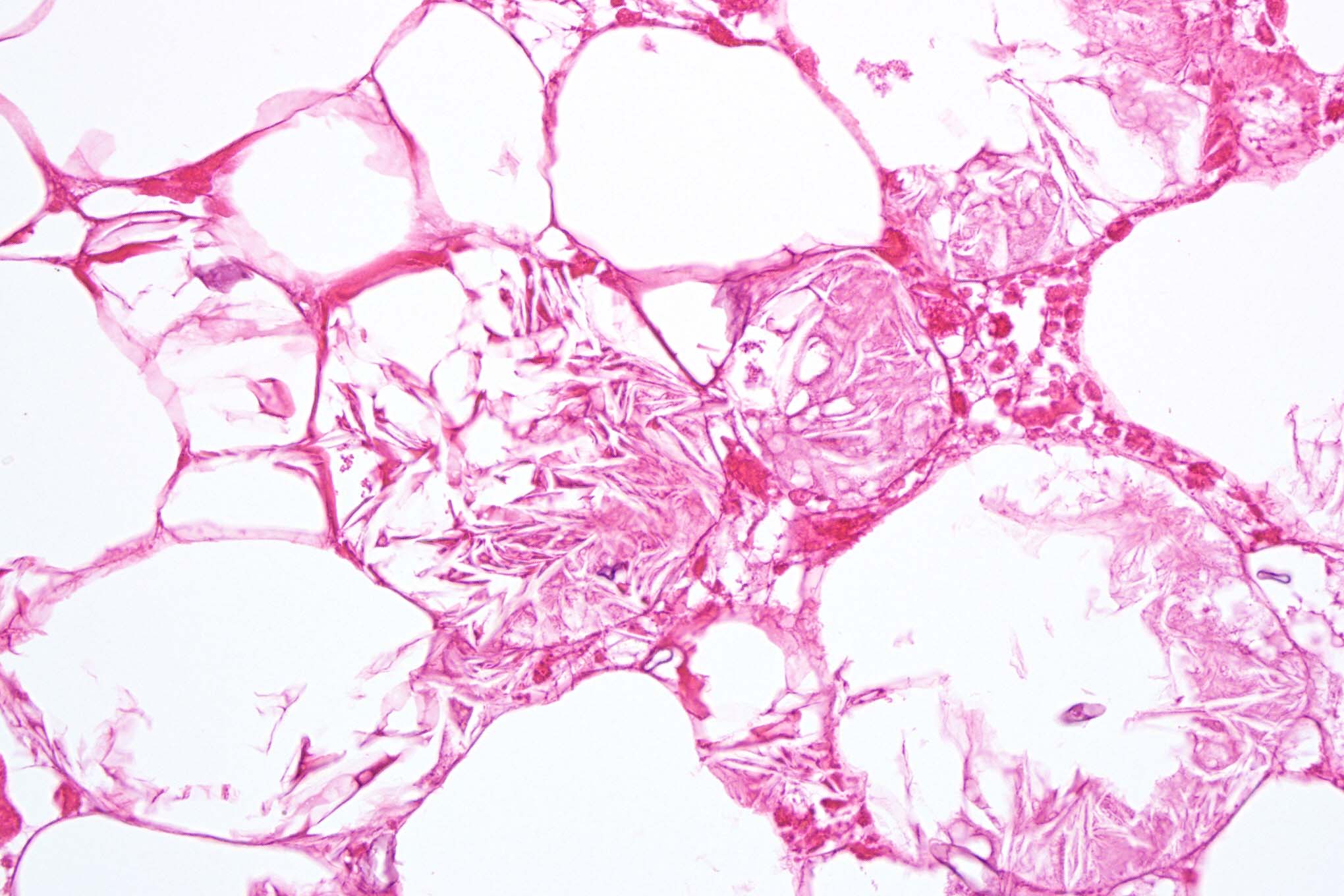
Male 42 years with deep nodule in thigh. Case courtesy of Dr. Thomas Brenn.


  Report Record

User Feedback


Guest Dr Engin Sezer

Posted

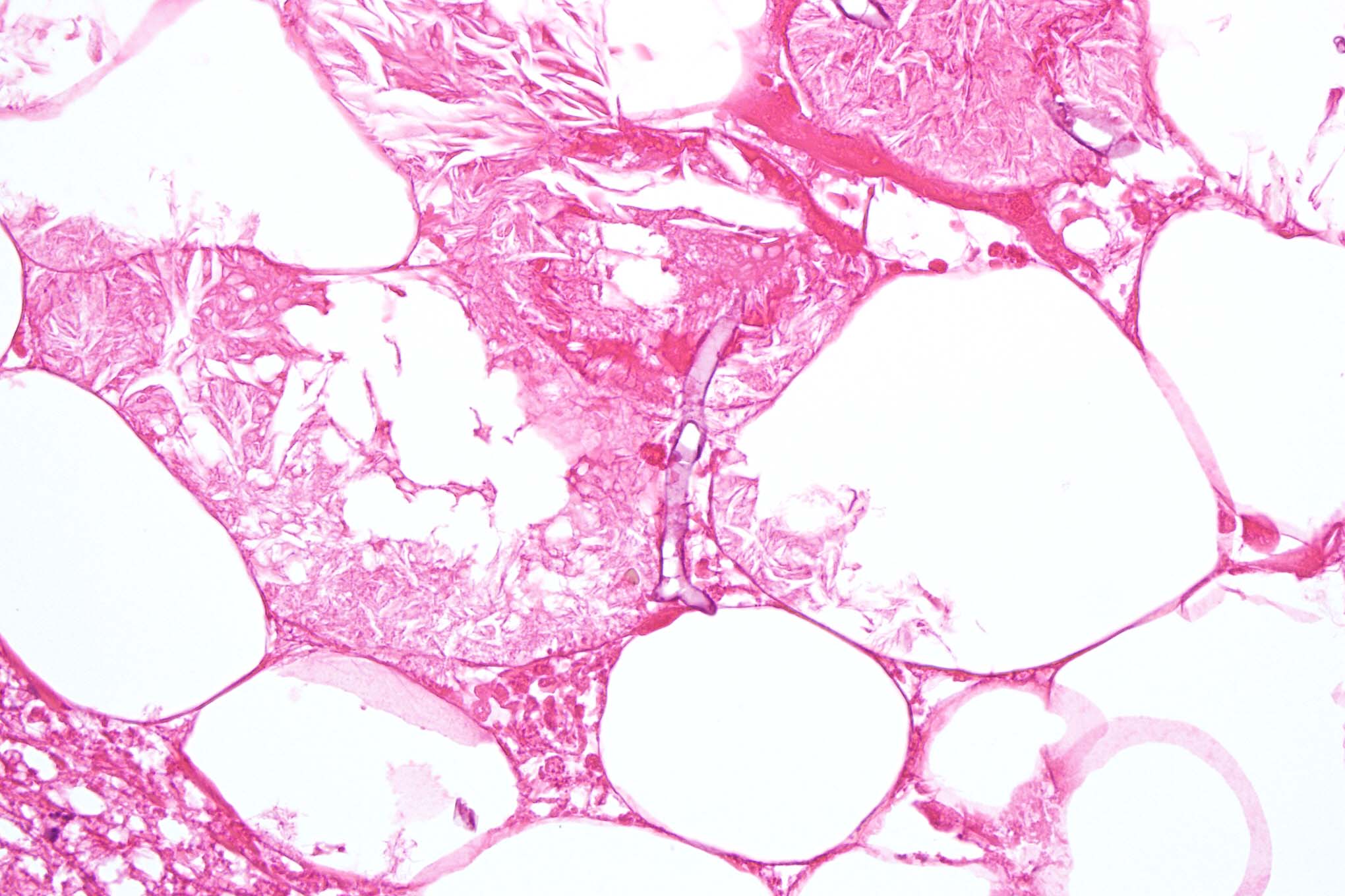
Aspergillosis vs. zygomycosis (favoring the preceding diagnosis based on septate appearance with 45 angle on fig. 4)

Share this comment


Link to comment
share_externally

Guest Marwa Fawzy

Posted

I can see necrosis of SC fat and the individual fat cells are swollen and contain abandant esinophilic radially arranged crystalline spaces with heavy infiltrate of esinophils and histiocytes .this picture can be seen in SCfat necrosis and post steroid panniculitis which has recently reported in adult patient.

Share this comment


Link to comment
share_externally

Dr. Mona Abdel-Halim

Posted

I think the fungal structure in the last image is broad non septate. This will favor Zygomycosis over Aspergillosis. And the extensive necrosis of the fat will indicate angioinvasion, so I will go for Mucormycosis.

Share this comment


Link to comment
share_externally

Dermatophtyic infection, ?zygomycosis.

Share this comment


Link to comment
share_externally

Sasi Attili

Posted

[quote name='Dr. Mona Abdel Halim' timestamp='1355229700']
I think the fungal structure in the last image is broad non septate. This will favor Zygomycosis over Aspergillosis. And the extensive necrosis of the fat will indicate angioinvasion, so I will go for Mucormycosis.
[/quote]

agree

Share this comment


Link to comment
share_externally

Guest Robledo F. Rocha

Posted

[color="#000000"][font="Palatino Linotype, serif"][size="3"]I favor the diagnosis of mucormycosis. There's a paucicellular necrotizing panniculitis with numerous broad hyphae that appear as negative shadows. There's also a septal abscess rich in eosinophils surrounding a thrombosed vessel, a sign of fungal angioinvasion.[/size][/font][/color]

Share this comment


Link to comment
share_externally

Guest MarieMD

Posted

Zygomycosis...given that this is subcutaneous, I would consider Basidiobolus.

Share this comment


Link to comment
share_externally

Dr. Phillip McKee

Posted

You are all very good. The fungus is a member of the Zygomycosis species. I do not have any further information.

Share this comment


Link to comment
share_externally

Arif Usmani

Posted

Marwa mentioned crystalline structure which in my limited experience is not typical for infectious panniculitides. However this paper mentions presence of needle-shaped crystals in a study of 13 case of mucormycosis subcutaneous infection.

[b] [b][font=arial,helvetica,sans-serif]Cutaneous and mucosal mucormycosis mimicking pancreatic [size=4]panniculitis[/size] and gouty [size=4]panniculitis[/size].[/font][/b][/b]

[b][font=arial,helvetica,sans-serif][url="http://www.ncbi.nlm.nih.gov/pubmed?term=eosinophilic%20panniculitis%2C%20hyphae%2C#"]J Am Acad Dermatol.[/url] 2012 Jun;66(6):975-84. [/font][/b]

Share this comment


Link to comment
share_externally


×
×
  • Create New...