Jump to content
In this section we have spot diagnoses posted on a daily basis since June 2010, now over 4000! You can review the archived cases and read the suggested diagnoses by users and the final comment by the contributors.
Case are uploaded each week day by 10 am UK time with the correct diagnosis will generally be posted at 8 pm UK time. Why not view the most recent spot diagnosis and proffer a diagnosis?

Case Number : Case 656 - 13 Dec Posted By: Guest

Please read the clinical history and view the images by clicking on them before you proffer your diagnosis.
Submitted Date :
   (0 reviews)

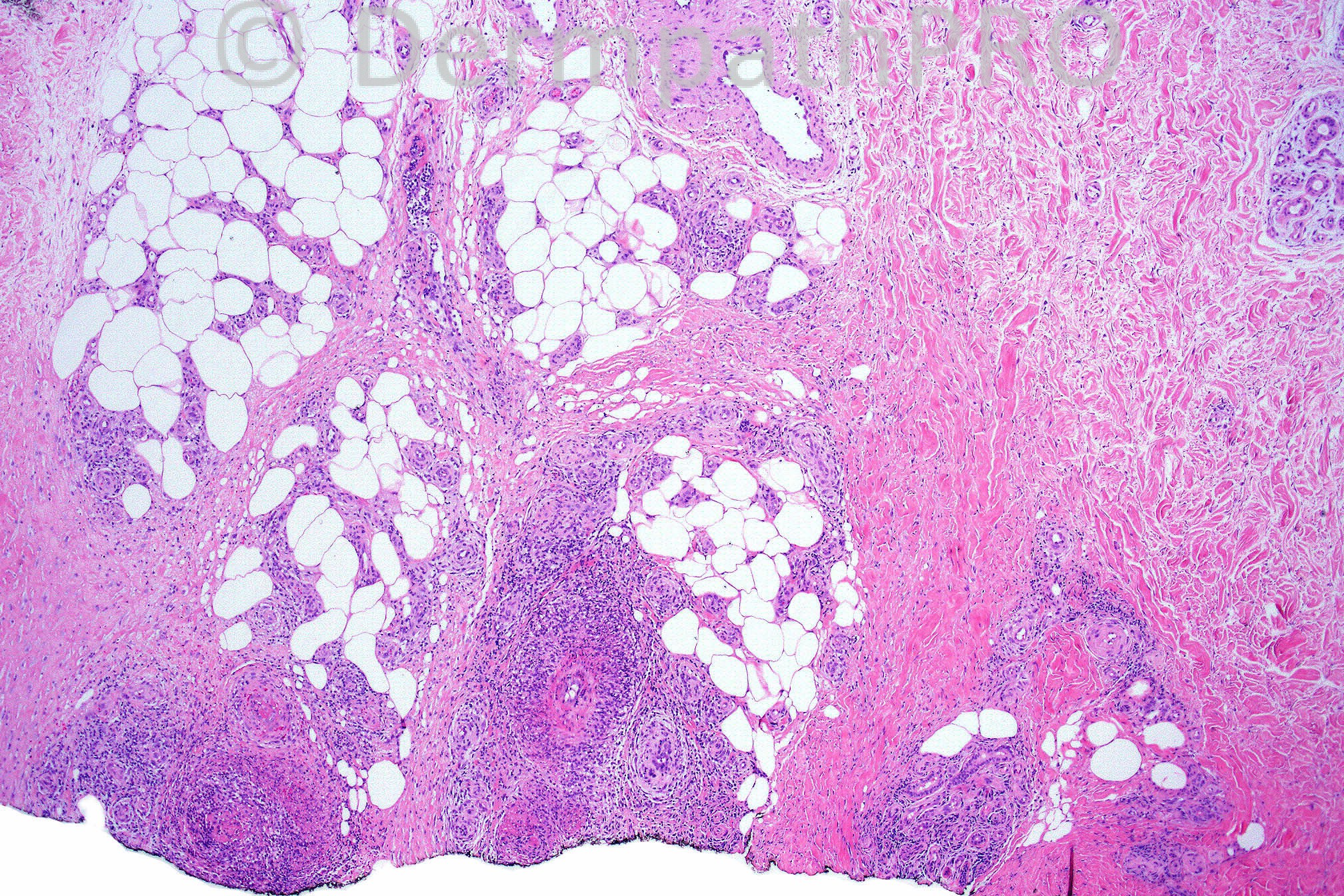
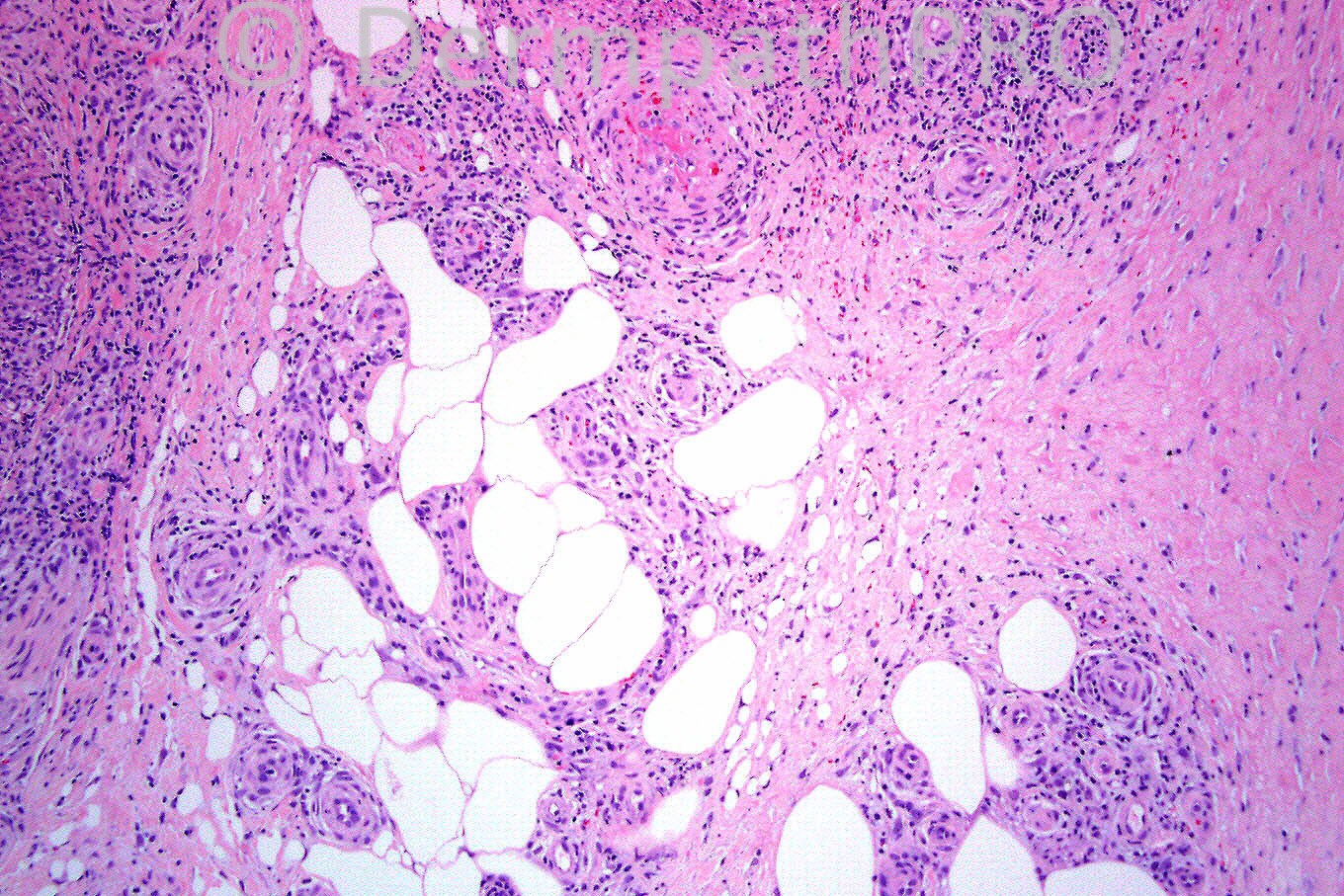
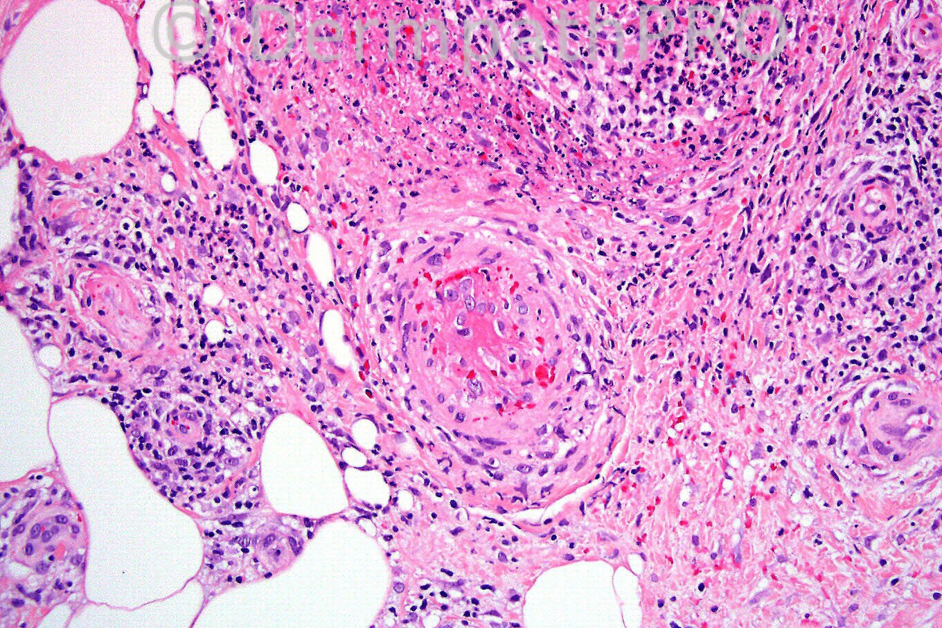
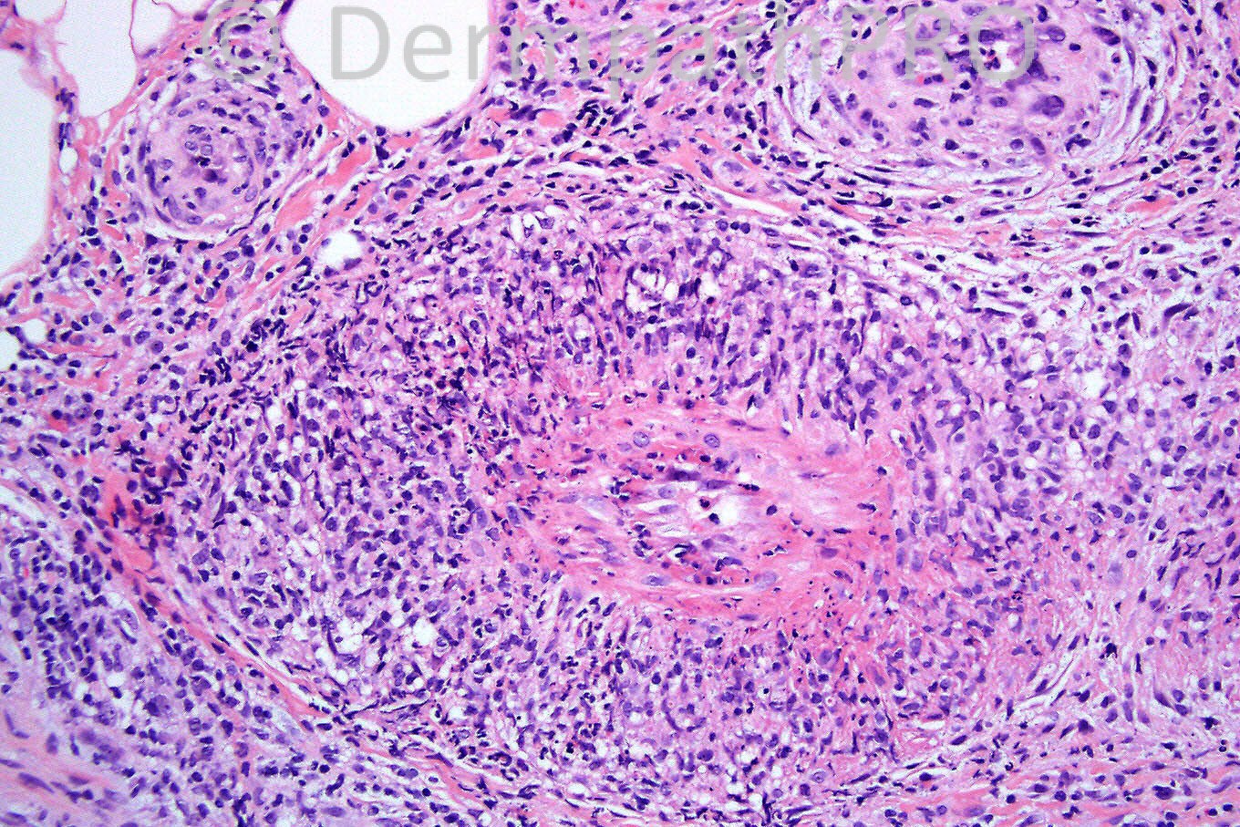
Female 27 years with tender nodules on calves. Case courtesy of Dr. Vince Liu.


  Report Record

User Feedback


Guest Hazem Hamed

Posted

Mixed lobular and septal panniculitis due to vasculitis. I would favor polyarteritis nodosa for correlation with serology.

Share this comment


Link to comment
share_externally

Agree with Dr. Saeb DD, however I would prefer erythema induratum of Bazin.

Share this comment


Link to comment
share_externally

Dr. Mona Abdel-Halim

Posted

Favor Erythema Induratum of Basin (nodular vasculitis).

Share this comment


Link to comment
share_externally

Guest Robledo F. Rocha

Posted

I think it’s a case of classic polyarteritis nodosa. There’s a leukocytoclastic vasculitis of small and medium-sized arteries of the subcutaneous fat. Typically, damaged vessels are in different stages of development. Early lesion (right bottom) shows fibrinoid necrosis of the arterial wall with a mixed infiltrate rich in neutrophilic nuclear dust present within and around the arterial wall. Late lesion (left bottom) is evidenced by perivascular fibrosis and occlusion of the vessel lumen by myxoid fibro-intimal proliferation.
As the resulting panniculitis seems to be restricted to the vicinity of the involved vessels and granulomas couldn’t be found, I suppose less likely the diagnosis of erythema induratum / nodular vasculitis.

Share this comment


Link to comment
share_externally

Sasi Attili

Posted

The inflammation seems to be mixed but predominantly septal. I do not think vasculitis in any way rules out Erythema nodosum, which would be my favoured diagnosis in this case given the clinical. However I have found it difficult to distinguish this from vasculitis seen in PAN and haven't yet seen a convincing explanation to differentiate the two. Would be grateful of Prof. McKee can maybe explain the differentiating features.

Share this comment


Link to comment
share_externally

Guest MarieMD

Posted

PAN...nice images of neutrophilic vasculitis of musc. vessels with occlusion

Share this comment


Link to comment
share_externally

Dr. Phillip McKee

Posted

The clinical history is of major importance. A female with tender nodules on the calves is characteristic of nodular vasculitis (erythema induratum). In erythema nodosum, the lesions are on the shins. There is a mixed septal and lobular panniculitis with both acute and chronic inflammation. Granulomata are not evident but this may be a result of the small size of the biopsy specimen and they may have been present elsewhere in the lesion (not biopsied). I think that PAN is a very good differential diagnosis but clinically, I doubt very much that lesions would have been limited to the calves. Obviously laboratory investigations would have been necesary to make a diagnosis of PAN. I have no information as the case was shared with me as an example of classical nodular vascuilitis. I think that vasculitis is always a difficult diagnostic differential as the features are not specific and overlap many conditions. Often a full clincial and laboratory workup are necessary before a definitive diagnosis can be made.

Share this comment


Link to comment
share_externally


×
×
  • Create New...