Jump to content
In this section we have spot diagnoses posted on a daily basis since June 2010, now over 4000! You can review the archived cases and read the suggested diagnoses by users and the final comment by the contributors.
Case are uploaded each week day by 10 am UK time with the correct diagnosis will generally be posted at 8 pm UK time. Why not view the most recent spot diagnosis and proffer a diagnosis?

Case Number : Case 657 - 14 Dec Posted By: Guest

Please read the clinical history and view the images by clicking on them before you proffer your diagnosis.
Submitted Date :
   (0 reviews)

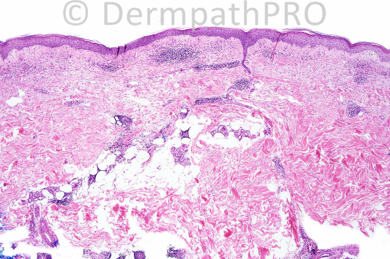
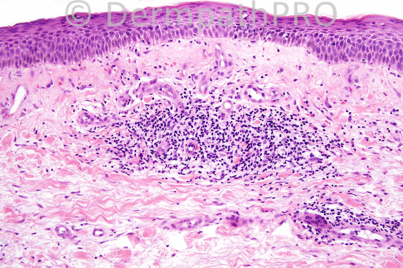
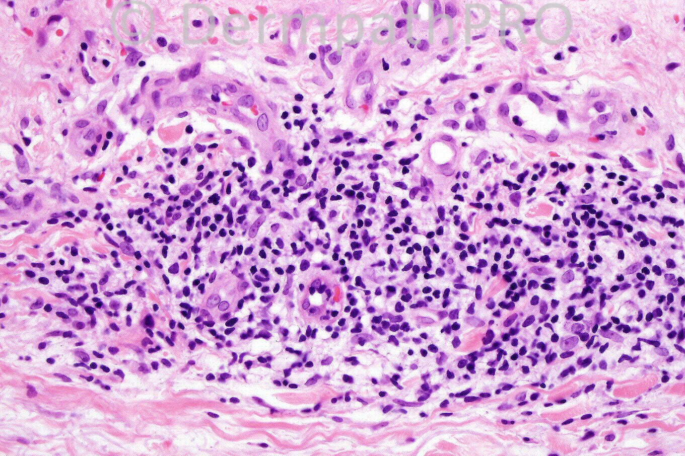
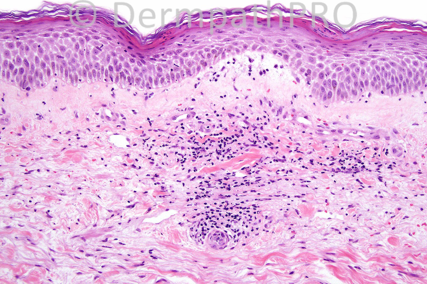
Female 26 years with erythematous urticarial lesions on limbs. Case courtesy of Dr. Vince


  Report Record

User Feedback


superficial and deep lymphocytic vasculitis which could correspond to urticarial vascultiis or end stage of other vasculitis maybe in a patient with SLE

Share this comment


Link to comment
share_externally

Guest Dr Engin Sezer

Posted

Lymhoid aggregates are present at the subcutaneous tissue as well. It would be nice to see the high magnification of the deeper tissue for lymphocytic dusts and hyalinizing necrosis for 'lupus panniculitis', which fits well with clinical presentation.

Share this comment


Link to comment
share_externally

Guest MarieMD

Posted

Superficial and deep perivascular dermatitis with spongiosis and papillar edema...I'm thinking of PMLE.
DDX: Mucha-haberman (?hints of interface in last image)

Share this comment


Link to comment
share_externally

Guest Robledo F. Rocha

Posted

I think that the best hypothesis is lupus erythematosus. Some clues of this disease are present, like sparse superficial and deep perivascular infiltrate of lymphocytes and some plasma cells with patchy inflammatory infiltrate in fat lobules, smudged appearance of the dermoepidermal interface, lamellar ortokeratosis alternates with parakeratosis in a checkerboard pattern, telangiectases in upper dermis, and, above all, blue deposits (mucin???) between strands of collagen. I couldn’t found any of the typical findings at the dermoepidermal junction, probably due to acuteness of the eruption expressed by some erythrocytes extravasated.

Share this comment


Link to comment
share_externally

Dr. Phillip McKee

Posted

Well done Marcela. The diagnosis is urticarial vasculitis. I don't know whether there was a background of lupus. The histological differential diagnosis includes any of the causes of superficial lymphocytic infitrates. The clinical history fits very well with the clincal diagnosis. Lupus profundus (panniculitis) presents with erythematous plaques, nodules and tumors and shows a predilection for the face trunk and limbs. Old lesions are seen as indurated depressed lesions.
The case was actually courtesy of Vince Liu.
Many thanks for all of your comments. Have a wonderful weekend.

Share this comment


Link to comment
share_externally


×
×
  • Create New...