Jump to content
In this section we have spot diagnoses posted on a daily basis since June 2010, now over 4000! You can review the archived cases and read the suggested diagnoses by users and the final comment by the contributors.
Case are uploaded each week day by 10 am UK time with the correct diagnosis will generally be posted at 8 pm UK time. Why not view the most recent spot diagnosis and proffer a diagnosis?

Case Number : Case 664 - 27 Dec Posted By: Guest

Please read the clinical history and view the images by clicking on them before you proffer your diagnosis.
Submitted Date :
   (0 reviews)

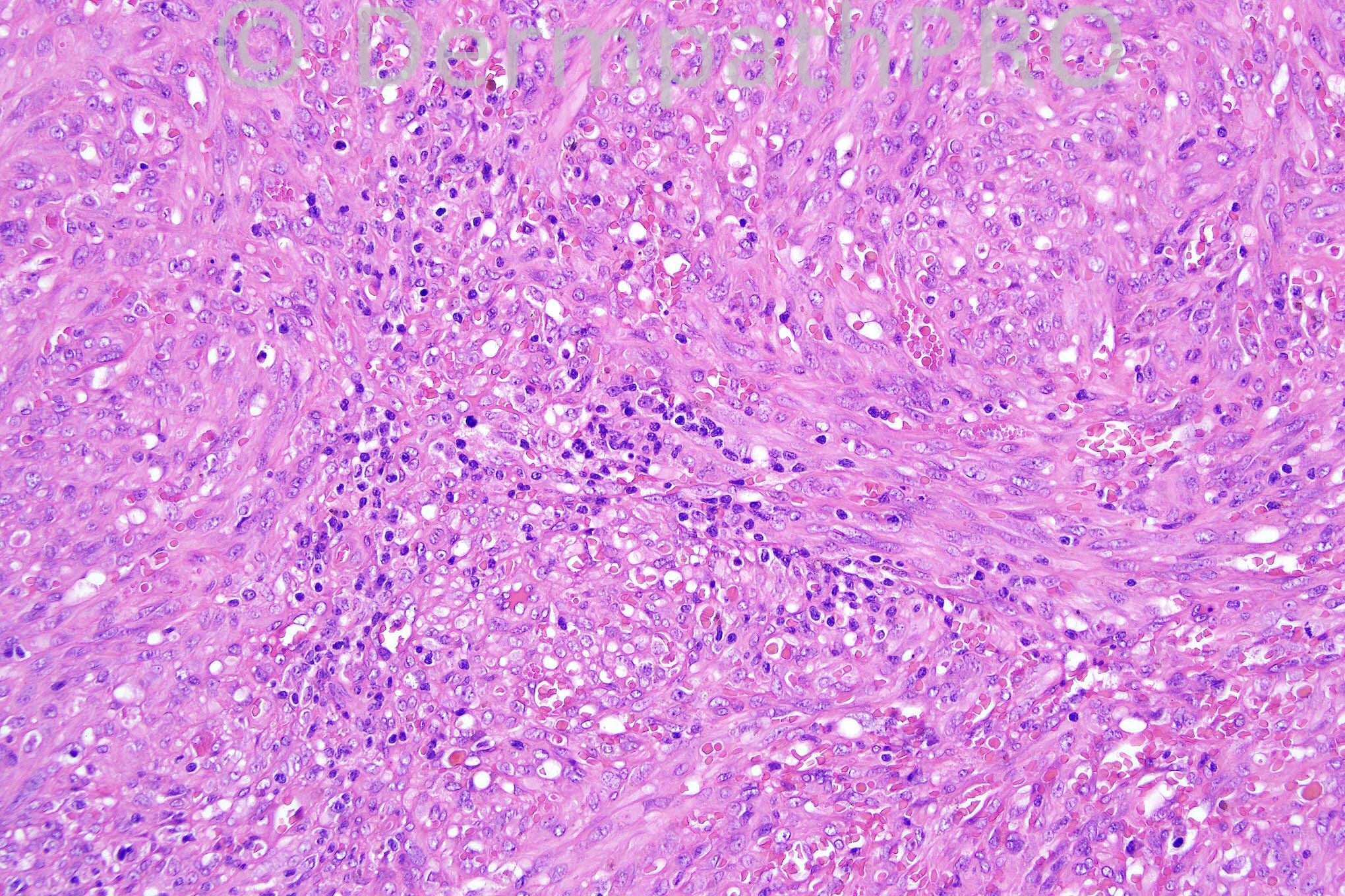
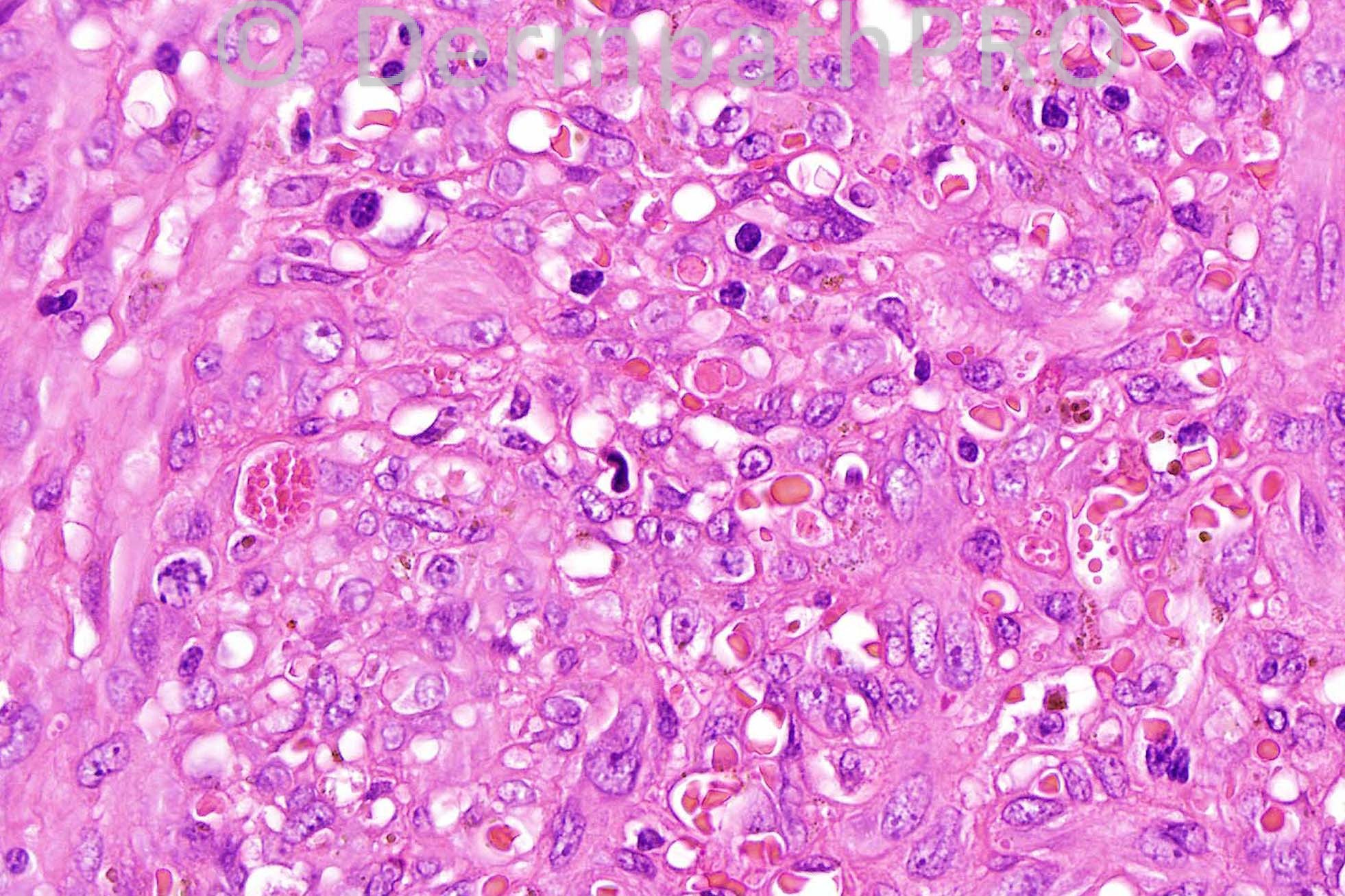
No clinical history. Courtesy of Wayne Grayson.


  Report Record

User Feedback


Guest Hazem Hamed

Posted

Kaposi's sarcoma for HHV8.

Share this comment


Link to comment
share_externally

Sasi Attili

Posted

What about Epithelioid Hemangioendothelioma?

Share this comment


Link to comment
share_externally

Guest Robledo F. Rocha

Posted

[font="'courier new"]Kaposi sarcoma, nodular stage. The dermis is expanded by a solid tumor made up of relatively monomorphic spindled cells laid out in fascicles, with slit-like vascular channels containing erythrocytes. There are large numbers of eosinophilic hyaline globules.[/font]

Share this comment


Link to comment
share_externally

Dr. Phillip McKee

Posted

Kaposi's sarcoma is correct. I thought that the eosinophilic hyaline globules were beautifully represented. Epithelioid hemangioendothelioma looks quite different. The tumor cells are dispersed in a myxoid and hyaline stroma and show intracytoplasmic lumina.

Share this comment


Link to comment
share_externally


×
×
  • Create New...