Jump to content
In this section we have spot diagnoses posted on a daily basis since June 2010, now over 4000! You can review the archived cases and read the suggested diagnoses by users and the final comment by the contributors.
Case are uploaded each week day by 10 am UK time with the correct diagnosis will generally be posted at 8 pm UK time. Why not view the most recent spot diagnosis and proffer a diagnosis?

Case Number : Case 441 Posted By: Guest

Please read the clinical history and view the images by clicking on them before you proffer your diagnosis.
Submitted Date :
   (0 reviews)

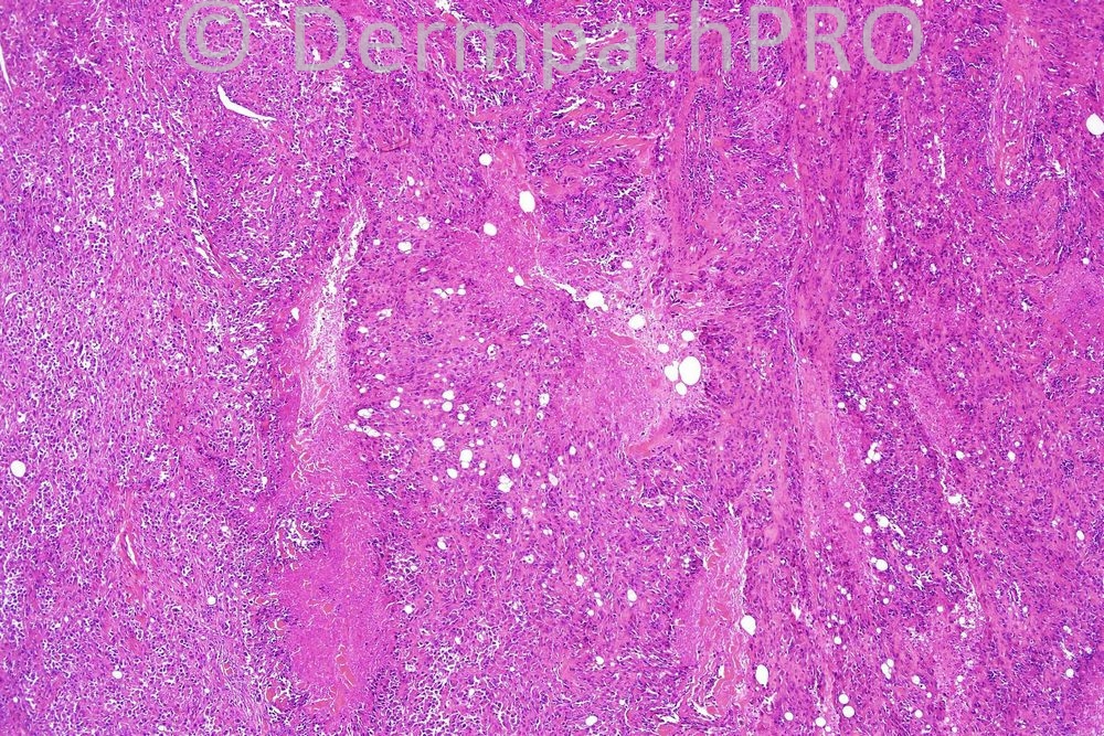
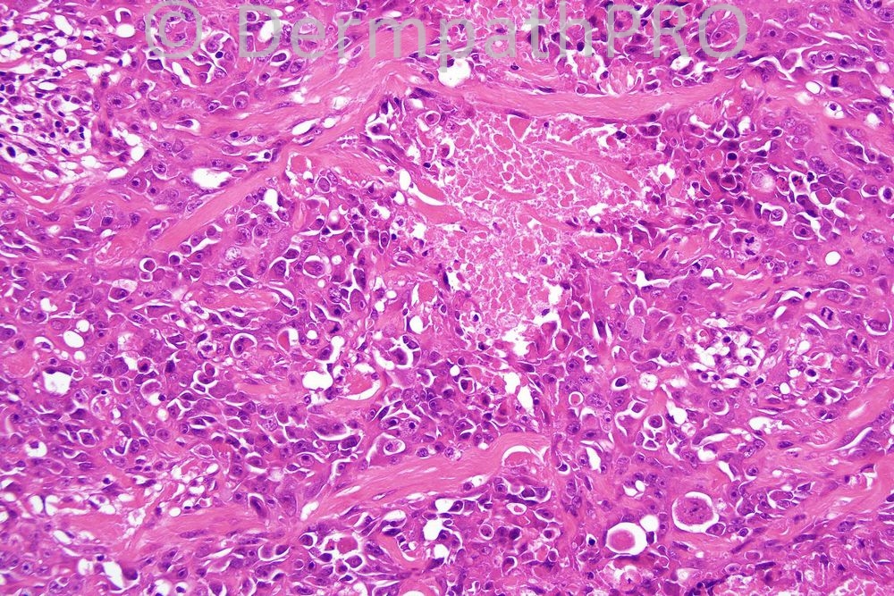
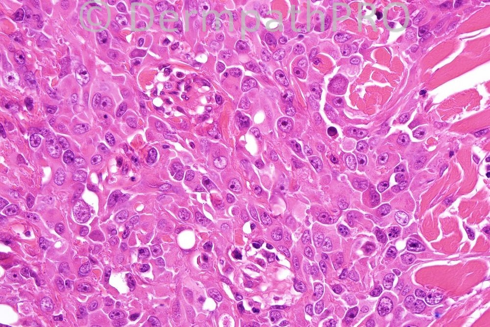
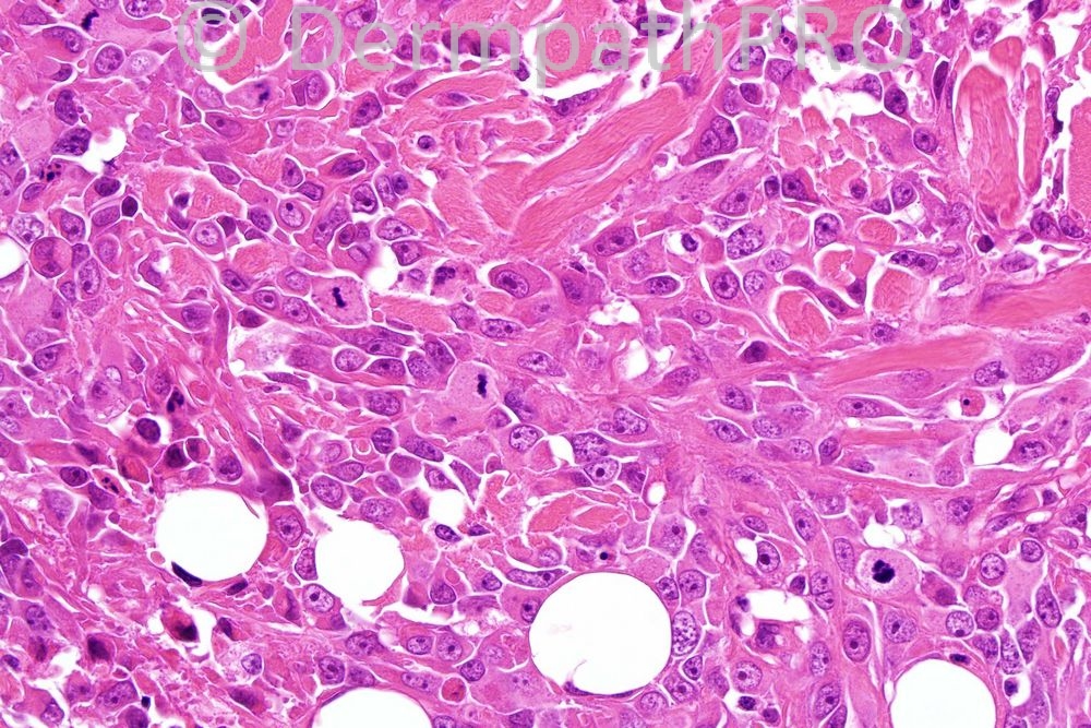
Male 81 years, ulcerated nodule on scalp.


  Report Record

User Feedback


Admin_Dermpath

Posted

[b]Francisco Vílchez - Department of Dermatology. Clinical Universitary Hospital "Virgen de la Victoria" (Málaga, Spain) Wrote:[/b]

Squamous cell carcinoma

[size=2]Submitted on 16/02/2012 16:51[/size] [b]Sasi Attili - (United Kingdom) Wrote:[/b]

I fin d this extremely difficult to diagnose based on H&E alone. I would have all the diagnoses mentioned below, in my differentials

[size=2]Submitted on 16/02/2012 16:24[/size] [b]Phillip McKee - Central Manchester NHS Trust (Manchester, UK) Wrote:[/b]

Much better responses - would require immuno' but yes the diagnosis is Epithelioid angiosarcoma. Thanks for all your comments.

[size=2]Submitted on 16/02/2012 16:22[/size] [b]Eman El-Nabarawy - Cairo University (Egypt) Wrote:[/b]

Melanoma vs epithelioid angiosarcoma.

[size=2]Submitted on 16/02/2012 15:03[/size] [b]Revelles - Fundación Jiménez Díaz (Madrid) Wrote:[/b]

I would go for epitheloid angiosarcoma

[size=2]Submitted on 16/02/2012 14:40[/size] [b]Richard Carr - Warwick (UK) Wrote:[/b]

Clinical would have made me go for a poorly differentiated acantholytic SCC first, melanoma second then rule out rarities like epitheliloid angiosarcoma, epithelioid sarcoma (central/high grade) and metastases

[size=2]Submitted on 16/02/2012 14:21[/size] [b]A Bansal - BCU HB (North Wales) Wrote:[/b]

Morphological impression: Epithelioid Angiosarcoma.

[size=2]Submitted on 16/02/2012 14:02[/size] [b]Engin Sezer - Department of Dermatology, Acıbadem Univ. (Istanbul, Turkey) Wrote:[/b]

Dissection of collagen in the lower portion of low magnification photomicrograph supports angiosarcoma histologically. I'm so curious regarding the immunos.

[size=2]Submitted on 16/02/2012 13:27[/size] [b]ANDREW DANCKWERTS - WITS UNIVERSITY, JOHANNESBURG (JOHANNESBURG, RSA) Wrote:[/b]

HIGH GRADE MALIGNANT NEOPLASM WITH AN EPITHELIOD MORPHOLOGY. IN VIEW OF THE MORPHOLOGY AND THE CLINICAL SCENARIO (ie. AGE OF PATIENT AND SITE OF LESION), MY DIFFERENTIAL DIAGNOSIS RESTS BETWEEN EPITHELIOD ANGIOSARCOMA (FAVOURED) AND AMELANOTIC MELANOMA. HOWEVER, ADDITIONAL DIFFERENTIAL DIAGNOSES INCLUDE THE FOLLOWING: METASTATIC LARGE CELL ADENOCARCINOMA AND MALIGNANT SKIN APPENDAGE TUMOUR, SUCH AS POROCARCINOMA. PAS / DIASTASE SPECIAL STAINS AND IMMUNOHISTOCHEMISTRY ARE ESSENTIAL TO DIFFERENTIATE.

[size=2]Submitted on 16/02/2012 12:54[/size] [b]José Leonardo Tovar - (BOGOTA-COLOMBIA) Wrote:[/b]

MELANOMA

[size=2]Submitted on 16/02/2012 12:14[/size] [b]Mona Abdel Halim - Dermatology Dept. Cairo Univ. (Egypt) Wrote:[/b]

My first impression is melanoma,,

[size=2]Submitted on 16/02/2012 11:34[/size] [b]Yüksel Okumuş - Bursa State Hospital (Turkey) Wrote:[/b]

Why not MELANOMA ?

[size=2]Submitted on 16/02/2012 10:03[/size] [b]Engin Sezer - Dermatology Department, Acıbadem Univ. (Istanbul, Turkey) Wrote:[/b]

Porocarcinoma vs. epithelioid angiosarcoma vs. poorly differeniated SCC. Panel including EMA, CD31 and MNF-116 may be helpful at this point.

[size=2]Submitted on 16/02/2012 09:58[/size] [b]Izzat Abdul-kadir - ST2 - York Hospital (UK) Wrote:[/b]

My immediate impression is sebaceous carcinoma.

[size=2]Submitted on 16/02/2012 09:53[/size]

Share this comment


Link to comment
share_externally


×
×
  • Create New...