Jump to content
In this section we have spot diagnoses posted on a daily basis since June 2010, now over 4000! You can review the archived cases and read the suggested diagnoses by users and the final comment by the contributors.
Case are uploaded each week day by 10 am UK time with the correct diagnosis will generally be posted at 8 pm UK time. Why not view the most recent spot diagnosis and proffer a diagnosis?

Case Number : Case 410 Posted By: Guest

Please read the clinical history and view the images by clicking on them before you proffer your diagnosis.
Submitted Date :
   (0 reviews)

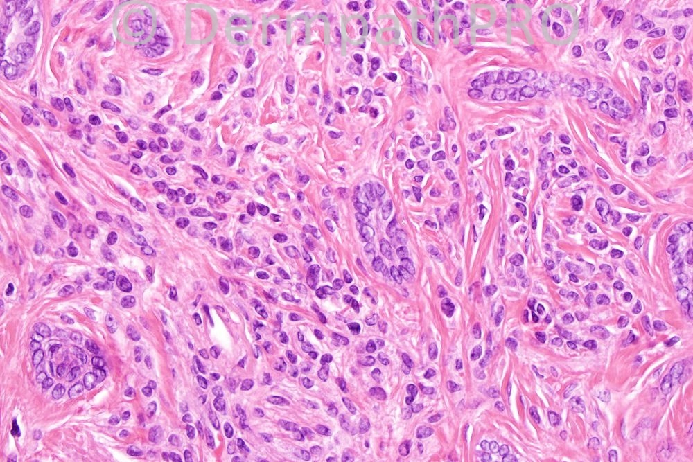
64 year old female, lesion left cheek, clinically dermal nevus with BCC.


  Report Record

User Feedback


Admin_Dermpath

Posted

Zena Slim (SpR Histopath) - () Wrote:

Thanks Abdul- Kadir. I didnt quite appreciate the basaloid nature of the cords across the screen

Submitted on 06/01/2012 22:29
________________________________________

carlos - Hosp miguel servet (spain) Wrote:

nevus + syringoma

Submitted on 05/01/2012 03:54
________________________________________

I. Abdul-kadir - Harrogate District Hospital (UK) Wrote:

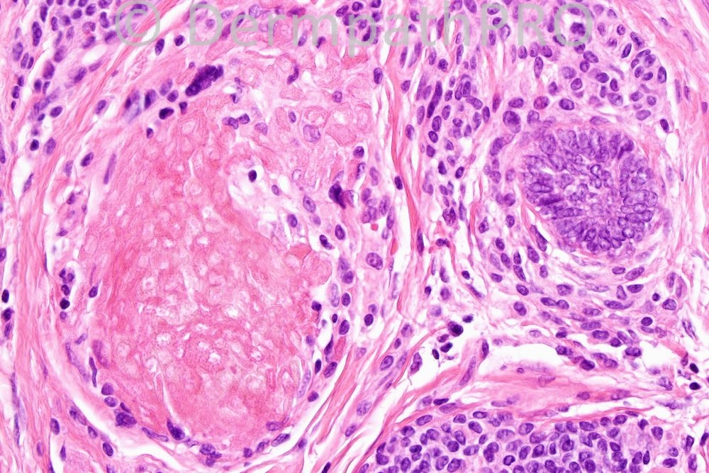
The trichepithelioma is the basaloid cells forming cords encircled by collaged and separated from one another by clefts (picture 2 & 3). The ghost cells & germ (picture 4) are signs of follicular differentiation.

Submitted on 05/01/2012 01:17
________________________________________

Zena Slim (SpR Histopath) - Wessex Deanery () Wrote:

I failed to see the Trichepitheliomatous component, please could you point me to which image shows it best? I thought that this was a benign intradermal naevus showing prominent features of 'maturation/senescence': fibrosis, calcification, ghost cells, etc.

Submitted on 04/01/2012 23:54
________________________________________

Phillip McKee - Overseas Consultations (Cave Creek, Arizona, USA) Wrote:

You are all correct. There is also a focus of ghost cells which I have not seen before in this lesion.

Submitted on 04/01/2012 22:18
________________________________________

Richard Carr - Wawick (UK) Wrote:

Akerman stated that DTE and naevus were a common association. We have seen the combination only once in around 20 cases of DTE (I keep a spreadsheet). Akerman also included the following comment: Shadow cells in cysts "often". We did not see it in any of our cases to date. Regards to all.

Submitted on 04/01/2012 22:16
________________________________________

Mona Abdel Halim - () Wrote:

Intradermal nevus and desmoplastic trichoepithelioma

Submitted on 04/01/2012 20:35
________________________________________

A Bansal - BCU HB (North Wales) Wrote:

Combined desmoplastic trichoepithelioma and intradermal nevus.

Submitted on 04/01/2012 18:32
________________________________________

Marcela Saeb Lima - INCMNSZ (Mexico City) Wrote:

intradermal nevus and desmoplastic trichoepithelioma with dystrophic calcification

Submitted on 04/01/2012 18:00
________________________________________

I. Abdul-kadir - Harrogate District Hospital (UK) Wrote:

Is it a combined desmoplastic trichoepithelioma and intradermal naevus?

Submitted on 04/01/2012 15:48
________________________________________

Share this comment


Link to comment
share_externally


×
×
  • Create New...