Jump to content
In this section we have spot diagnoses posted on a daily basis since June 2010, now over 4000! You can review the archived cases and read the suggested diagnoses by users and the final comment by the contributors.
Case are uploaded each week day by 10 am UK time with the correct diagnosis will generally be posted at 8 pm UK time. Why not view the most recent spot diagnosis and proffer a diagnosis?

Case Number : Case 542 - 6 July Posted By: Guest

Please read the clinical history and view the images by clicking on them before you proffer your diagnosis.
Submitted Date :
   (0 reviews)

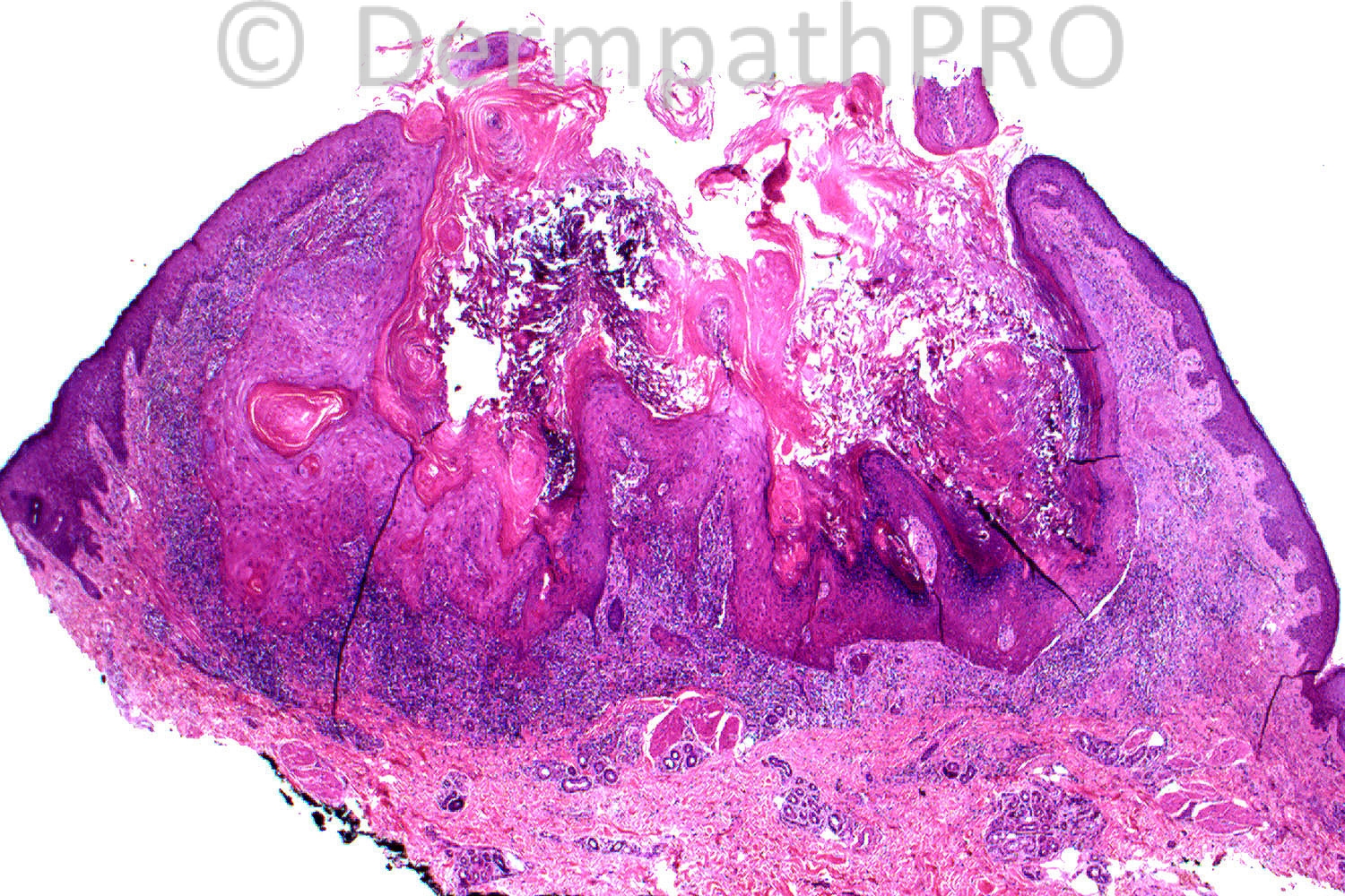
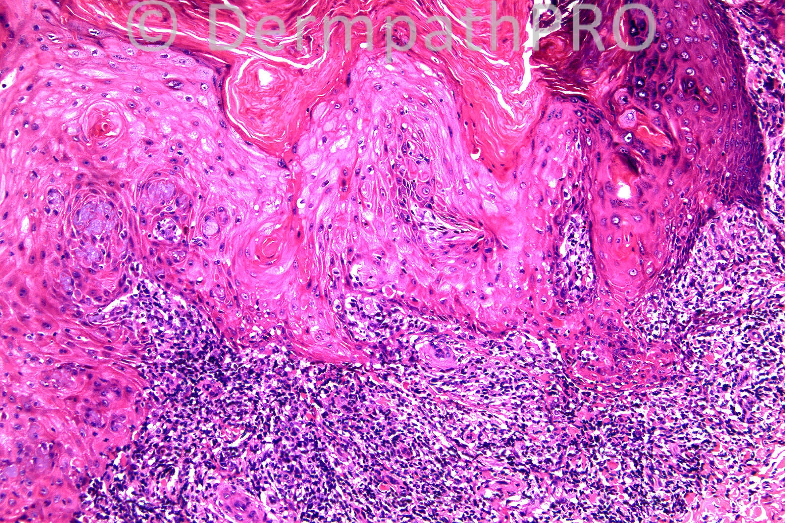
Female 70 years, arm, Nodular skin lesion L lower forearm ?BCC.


  Report Record

User Feedback


Admin_Dermpath

Posted

[b]Amr Gohar - Gohar's Skin and Sexual Health Clinic (Egypt) Wrote:[/b]

Well, as a clinician, certain features help me diagnose keratoacanthoma rather than SCC such as the description of the lesion, history of rapid growth, the undamaged surrounding skin and the younger age of onset and spontaneous regression, hence the importance of clinicopathological correlation. When there is evidence of actinic damage the clinical distinction is made more difficult. Pathologically, many features favour a diagnosis of keratoacanthoma over SCC ( the low-power architecture - lack of anaplasia - presence of intra-epithelial elastic fibres and intra-cytoplasmic glycogen - markers are just examples) however, using the small punch biopsies in elderly patients a superimposed squamous cell carcinoma might not be excluded hence the importance of studying both architecture and the cellular characteristics including the base and the edges and finally because SCC may masquerade as keratoacanthoma clinically and pathologically the term "keratoacanthoma-like SCC" is used at times, considering also the concept of malignant transformation.

[size=2]Submitted on 07/07/2012 12:46[/size]
[b]I. Abdul-kadir (ST2 histopath) - York Hospital (UK) Wrote:[/b]

This paper concludes that intraepithelial elastic fibers, among others, "were not distinctive criteria (of KA from SCC), although they are considered as classic distinctive features". Cribier B, et al. Differentiating squamous cell carcinoma from keratoacanthoma using histopathological criteria: Is it possible? A study of 296 cases. Dermatology 1999;199:208–212.

[size=2]Submitted on 07/07/2012 07:04[/size]
[b]Amr Gohar - Gohar's Skin and Sexual Health Clinic (Egypt) Wrote:[/b]

The presence of intra-epithelial elastic fibres has been said to be one of the features that favour the diagnosis of keratoacanthoma rather than SCC.

[size=2]Submitted on 07/07/2012 03:08[/size]
[b]Richard Carr - Warwick (UK) Wrote:[/b]

Yes the is a regressing KA. When the lesions is regressed to this extent but has intra-epithelial elastic fibres as a clue to the prior rapidity of growth there really is no differential diagnosis. Certainly I have never seen inverted follicular keratosis with incorporated elastic like this. Regards to all and enjoy the weekend.

[size=2]Submitted on 06/07/2012 17:53[/size]
[b]Marie MD - () Wrote:[/b]

KA

[size=2]Submitted on 06/07/2012 16:17[/size]
[b]Phillip H McKee - Overseas Consultations (Cave Creek, AZ, United States) Wrote:[/b]

Please visit my Facebook page to read the comments I have made about the new website. www.dermathPRO.com I am sure that you will all love it!!! Best wishes Phillip

[size=2]Submitted on 06/07/2012 15:36[/size]
[b]I. Abdul-kadir (ST2 histopath) - York Hospital (UK) Wrote:[/b]

KA versus KA-like SCC.

[size=2]Submitted on 06/07/2012 14:54[/size]
[b]Hazem Hamed - Imperial College (London) Wrote:[/b]

Thank you very much Richard. I missed it initially but yes transepithelial elimination of elastic fibers is frequently seen in keratoacanthoma.

[size=2]Submitted on 06/07/2012 13:02[/size]
[b]Richard Carr - Warwick (UK) Wrote:[/b]

Actually the clue I was referring to was the presence of elastic in the epithelium shown even better on the left side of the lower left image. The staining here is very blue and the elastic is actually a dull blue color. If you look carefully you can see blue elastotic material incorporated within the squamous epithelium.

[size=2]Submitted on 06/07/2012 12:13[/size]
[b]Mona Abdel Halim - () Wrote:[/b]

Evolving KA vs. KA like inverted follicular keratosis.

[size=2]Submitted on 06/07/2012 11:43[/size]
[b]Yüksel Okumuº - Bursa State Hospital (Turkey) Wrote:[/b]

Keratoacanthoma. DD: Lichen planus-like keratosis simulating keratoacanthoma.

[size=2]Submitted on 06/07/2012 11:33[/size]
[b]Francisco Vílchez-Márquez - Department of Dermatology. Ceuta Universitary Hospital. (Ceuta, Spain.) Wrote:[/b]

Keratoacanthoma

[size=2]Submitted on 06/07/2012 11:24[/size]
[b]Engin Sezer - (Istanbul) Wrote:[/b]

Interface tissue response to keratoacanthoma.

[size=2]Submitted on 06/07/2012 11:17[/size]
[b]Hazem Hamed - Imperial College (London) Wrote:[/b]

Thank you Richard. I think it is KA in its early growth stage (colloid bodies) rather than regression phase. There is no background LP.

[size=2]Submitted on 06/07/2012 10:55[/size]
[b]Amira Tawdy - Cairo University (Egypt) Wrote:[/b]

KA

[size=2]Submitted on 06/07/2012 10:40[/size]
[b]Vítor Carneiro - Hospital Ponta Delgada (Azores - Portugal) Wrote:[/b]

Keratoacanthoma.

[size=2]Submitted on 06/07/2012 10:29[/size]
[b]Richard Carr - Warwick (UK) Wrote:[/b]

Dear Hazem. There is a clue in the lower right image that will answer your question.

[size=2]Submitted on 06/07/2012 10:29[/size]
[b]Hazem Hamed - Imperial College (London) Wrote:[/b]

KA growth pattern. In addition, hypergranulosis is seen (1st and 3rd images.) Band-like infiltrate, colloid bodies and squqmatization of basal layer (4th image). I think it is regressing keratoacanthoma and possibly background of hypertrophic lichen planus. Was the lesion rapidly growing?.

[size=2]Submitted on 06/07/2012 10:22[/size]
[b]Iskander H. Chaudhry - Central Manchester Trust (Manchester, U.K.) Wrote:[/b]

Dear All, I am delighted to let you know that Dr Phillip McKee will be launching his new website on Monday 9th July where the spot diagnoses will be accessible from. We will post a link to the new website on this website on Monday morning. Many thanks for your contributions to this site.

[size=2]Submitted on 06/07/2012 09:00[/size]

Share this comment


Link to comment
share_externally

Guest Iskander

Posted

The New Website is Live now!

Share this comment


Link to comment
share_externally

Dr. Richard Carr

Posted

Thank you for the additional discussion of this case.
I could spend a very long time discussing KA v's SCC.
Firstly a serious caveat - I am not sure the diagnosis is safe in the hands of
experts let alone non-experts to be honest!
That said we in Warwick are (currently) firmly in the camp that KA is a distinct
Clinical-Pathological diagnosis.
Provided a lesion is typical clinically and histologically we can make a
confident diagnosis (in the knowledge that there is a risk of malignant
and exceedingly rarely metastasis usually to local lymph nodes).
Early KA (in our view) does often include quite striking cellular pleorphism
and typically the early lesions have highly proliferative infiltrative
(not pushing circumscript borders like well differentiated SCC) borders with
prominent elastic incorporation in the proliferative phase.
Despite the frighteningly worrying peripheral component in all areas of the
tumor there is central (often abprupt) transition to highly differentiated
and distincitive pilar keratinisation.
In excision biopsies the the symmetrical exoendophytic nature and rounded
silhouette are obviously helpful. In a small biopsy one can
easily diagnosis an early KA as a poorly (not well) differentiated SCC if one
does not spot the central differentiation in all islands (suggest multiple
levels are examined on small biopsies to demonstrate this feature of KA).
Incorporation of elastic can be a feature of poorly differentiated SCC but not,
in my experience a well differentiated SCC (that usually have much more pushing
mitotically inactive borders)and in my view is rarely a true diagnostic
consideration in the histological differential diagnosis of an early KA.
Obviously clinical information is essential as well differented SCC does not
grow rapidly in a few weeks like a KA.
Otherwise typical clinicopathological KA with frank (bowenoid) atypia
(probably about 30% of our cases of early KA!) in my opinion is of uncertain
biological significance but would lead us to recommend complete excision
(in partial biopsies i.e. malignant transformation must be a serious consideration)
and probably follow-up as for SCC (when seen in isolation in an excision specimen).
We tend to opt for complete excision with clear margins on most cases (of
even classicaly clinicopathological early KA) in any case. Often patients prefer
not to have an unsightly smelly rapidly growing lesion on their face and
conservative excision is cosmetically acceptable in good hands.
However for some typical (biopsy confirmed) cases watchful waiting (i.e. active
close follow-up with a low threshold for excision of persistent or actively
growing lesions) may be an option for some individual lesions for some patients.
Obviously patient's and clinicians will vary on how brave they are prepared to
be watching a lesion that may or may not undergo true malginant transformation.
Because of the complexity of the above and the many caveats you will hopefully
understand why I am saying I am not sure the diagnosis of KA is entirely safe!
I have not even discussed the many lesions we are now referring to as follicular
SCC that may closely, at first glance, resemble KA (often they have acantholysis
and central follicular mucin - features we do not see in KA - but most are well
differentiate with lobular circumscribed pushing bordres). We also often
sit on the fence or admit defeat in a significant proportion of cases (even
with good clinico-pathological corration).
We certainly discuss each and every case in a multi-disciplinary setting and
I hope an awareness of the difficulties and a shared team approach is the
safest way of managing these patient's.

Share this comment


Link to comment
share_externally


×
×
  • Create New...