Jump to content
In this section we have spot diagnoses posted on a daily basis since June 2010, now over 4000! You can review the archived cases and read the suggested diagnoses by users and the final comment by the contributors.
Case are uploaded each week day by 10 am UK time with the correct diagnosis will generally be posted at 8 pm UK time. Why not view the most recent spot diagnosis and proffer a diagnosis?

Case Number : Case 543 - 9 July Posted By: Guest

Please read the clinical history and view the images by clicking on them before you proffer your diagnosis.
Submitted Date :
   (0 reviews)

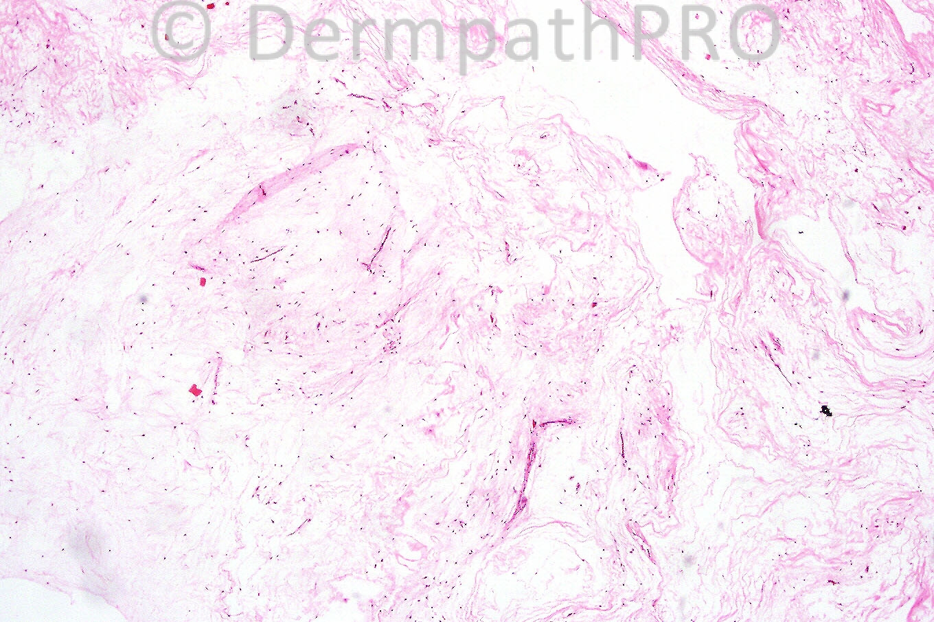
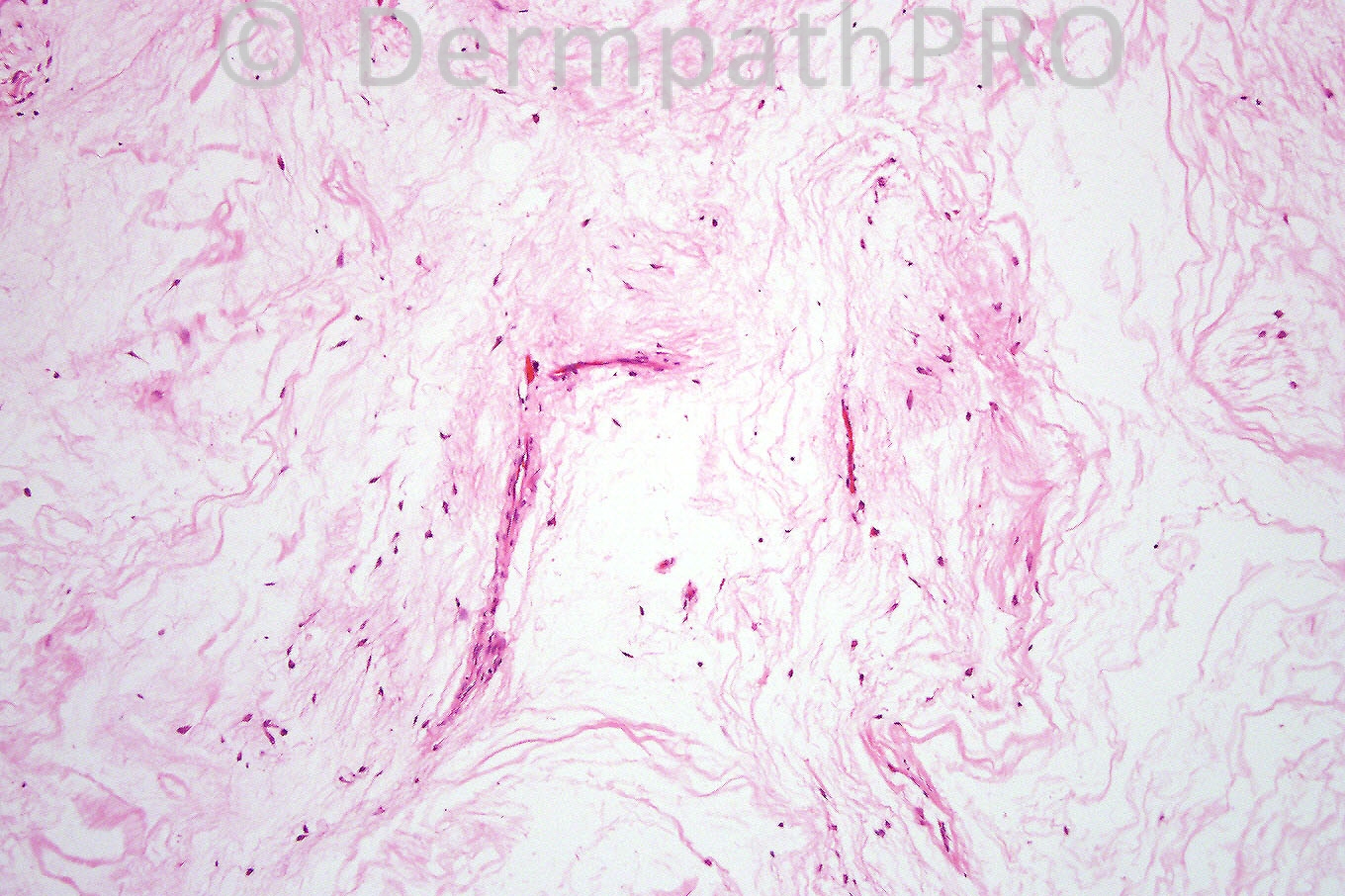
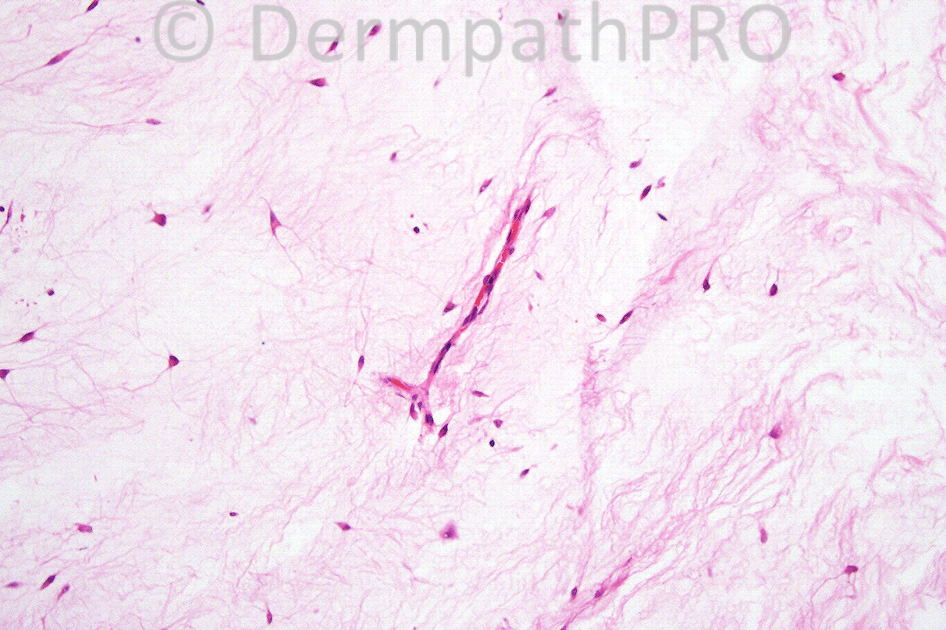
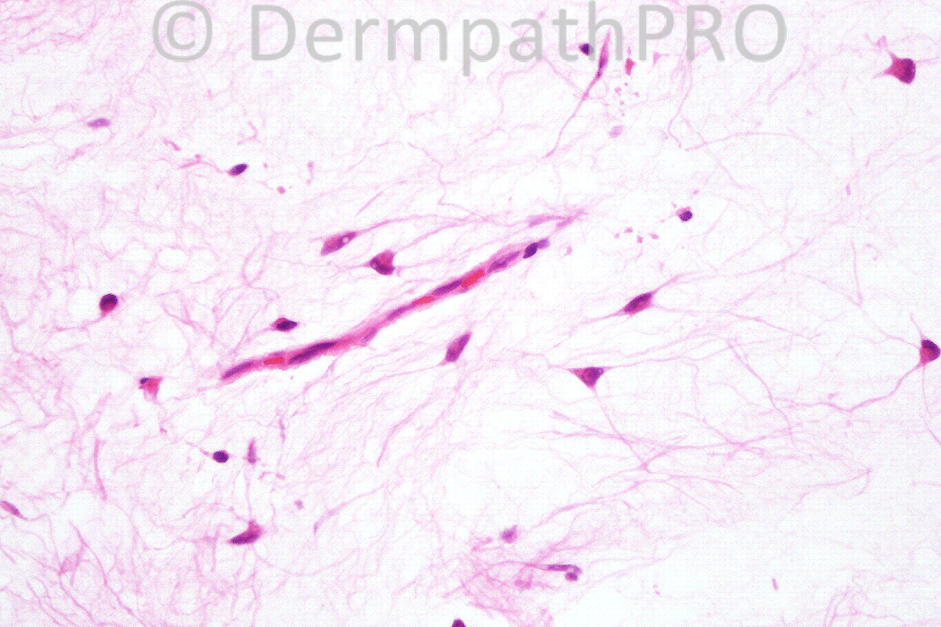
Female 82 years with a cystic swelling in the left groin.


  Report Record

User Feedback


Guest Hazem Hamed

Posted

Cutaneous myxoma. Is not richly vascular to call it angiomyxoma. Is there any manifestions of Carney's complex.

Share this comment


Link to comment
share_externally

Guest Francisco Vílchez-Márquez

Posted

Agree, myxoma. Congratulations for the new website!

Share this comment


Link to comment
share_externally

Guest Dr Engin Sezer

Posted

Myxoma, Prof. McKee many thanks and congratulations for this beautiful website

Share this comment


Link to comment
share_externally

Sasi Attili

Posted

Wonderful site Prof. McKee. I think the site (groin) is typical and the numerous blood vessels , stellate/spindle cells in the stroma, suggest an angiomyxoma

Share this comment


Link to comment
share_externally

Guest Juan Carlos Garcés

Posted

Cutaneous myxoma

Share this comment


Link to comment
share_externally

Dr. Phillip McKee

Posted

I included two images of vessels to draw your attention to the vascular component. I diagnosed this lesion as an angiomyxoma. I am unaware of any history of Carney's complex. Many thanks for your kind comments about the new website. We have been working towards this day ever since mckeedermpath.com was launched. It has taken about 4 years of very hard work to get here so it is a very exciting day for myself and Iskander.

Share this comment


Link to comment
share_externally

Guest El-Khalawany

Posted

Dear Prof. McKee,
A wonderful site, amazing cases and great effort.
Congratulation and all the best in the feature.
Mohamed El-Khalawany.

Share this comment


Link to comment
share_externally

Guest Amr Gohar

Posted

[font=courier new,courier,monospace][size=1][size=1][font="Revival565BT-Roman"][size="2"][font="Revival565BT-Roman"][size="2"][color=#222222]Cutaneous myxoma. There are thin collagen bundles and fibroblasts scattered through a mucinous matrix. Cutaneous myxomas are rare tumours [/color][color="#000000"]that may be associated with Carney’s complex. [color=black]Solitary and disseminated cutaneous myxomas unassociated with any systemic abnormalities may also occur.[/color][/color][/size][/font][/size][/font][/size][/size][/font]

Share this comment


Link to comment
share_externally

Guest Amr Gohar

Posted

[font=arial,helvetica,sans-serif][font=Revival565BT-Roman][size=6][font=Revival565BT-Roman][size=6][font=Revival565BT-Roman][size=6][font=Revival565BT-Roman][size=6]When the vascular component is marked, the term ‘angiomyxoma’ can be used. [/size][/font][/size][/font][/size][/font][/size][/font][/font]

Share this comment


Link to comment
share_externally

Dr. Richard Carr

Posted

I have added a blog on case 542 from July 6th 2012 - regards Richard

Share this comment


Link to comment
share_externally

Guest Vinoo Sekhri

Posted

Angiomyxoma: seeing one after a while!
The image quality is superb.

Share this comment


Link to comment
share_externally


×
×
  • Create New...