Jump to content
In this section we have spot diagnoses posted on a daily basis since June 2010, now over 4000! You can review the archived cases and read the suggested diagnoses by users and the final comment by the contributors.
Case are uploaded each week day by 10 am UK time with the correct diagnosis will generally be posted at 8 pm UK time. Why not view the most recent spot diagnosis and proffer a diagnosis?

Case Number : Case 544 - 10 July Posted By: Guest

Please read the clinical history and view the images by clicking on them before you proffer your diagnosis.
Submitted Date :
   (0 reviews)

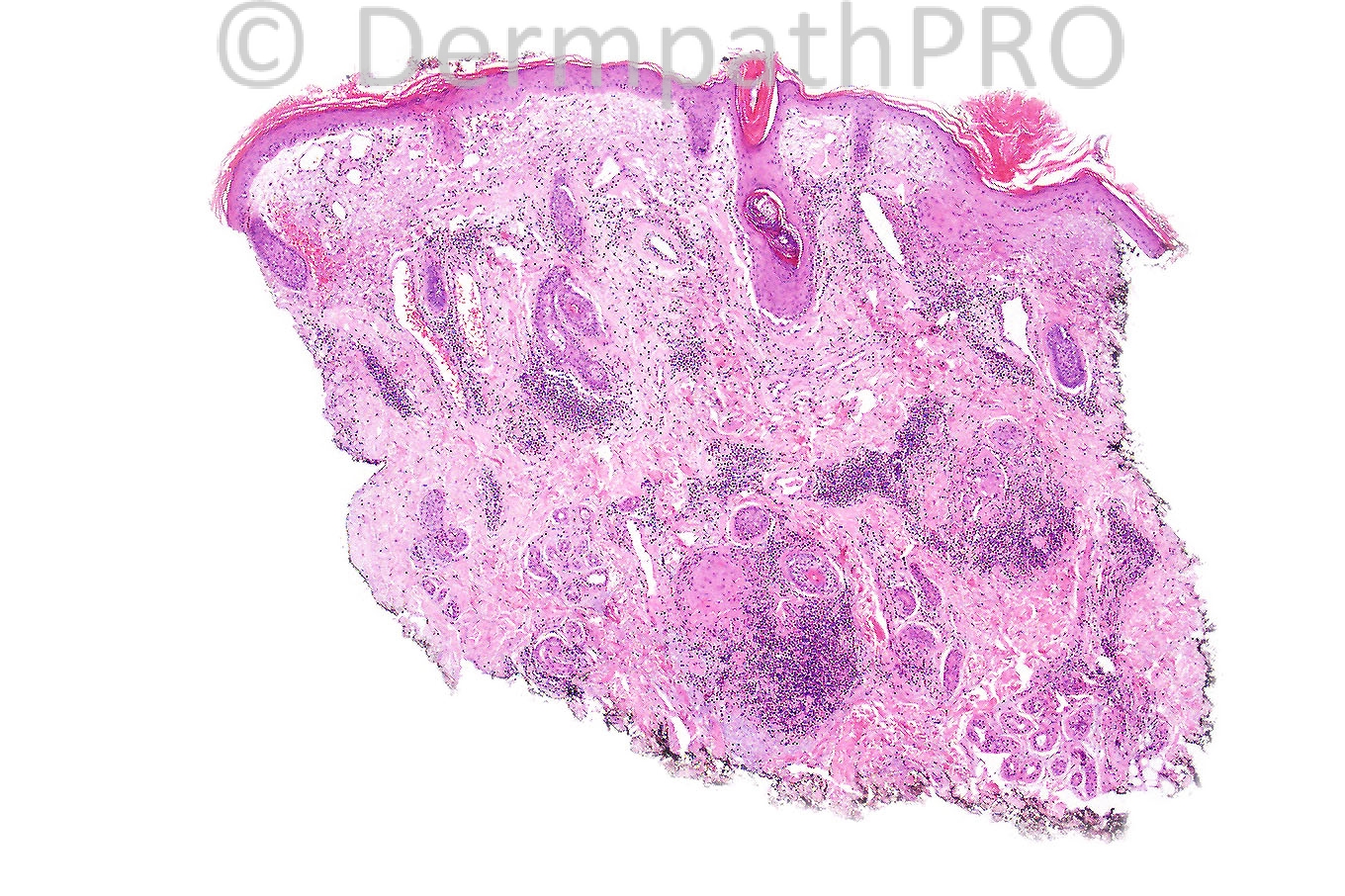
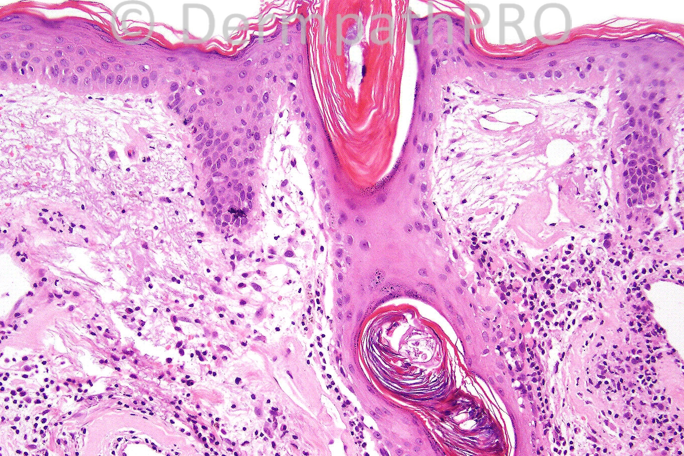
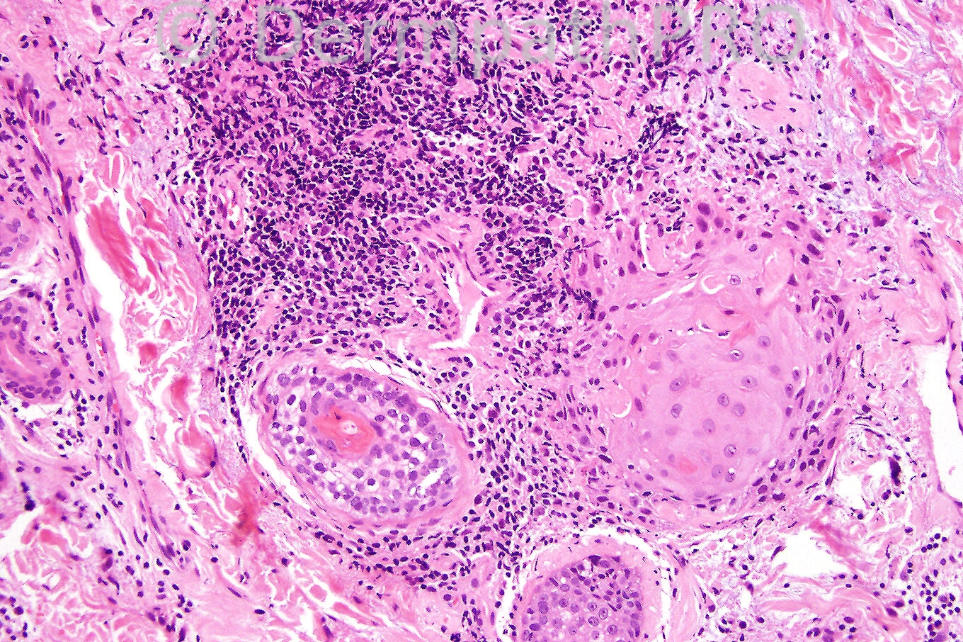
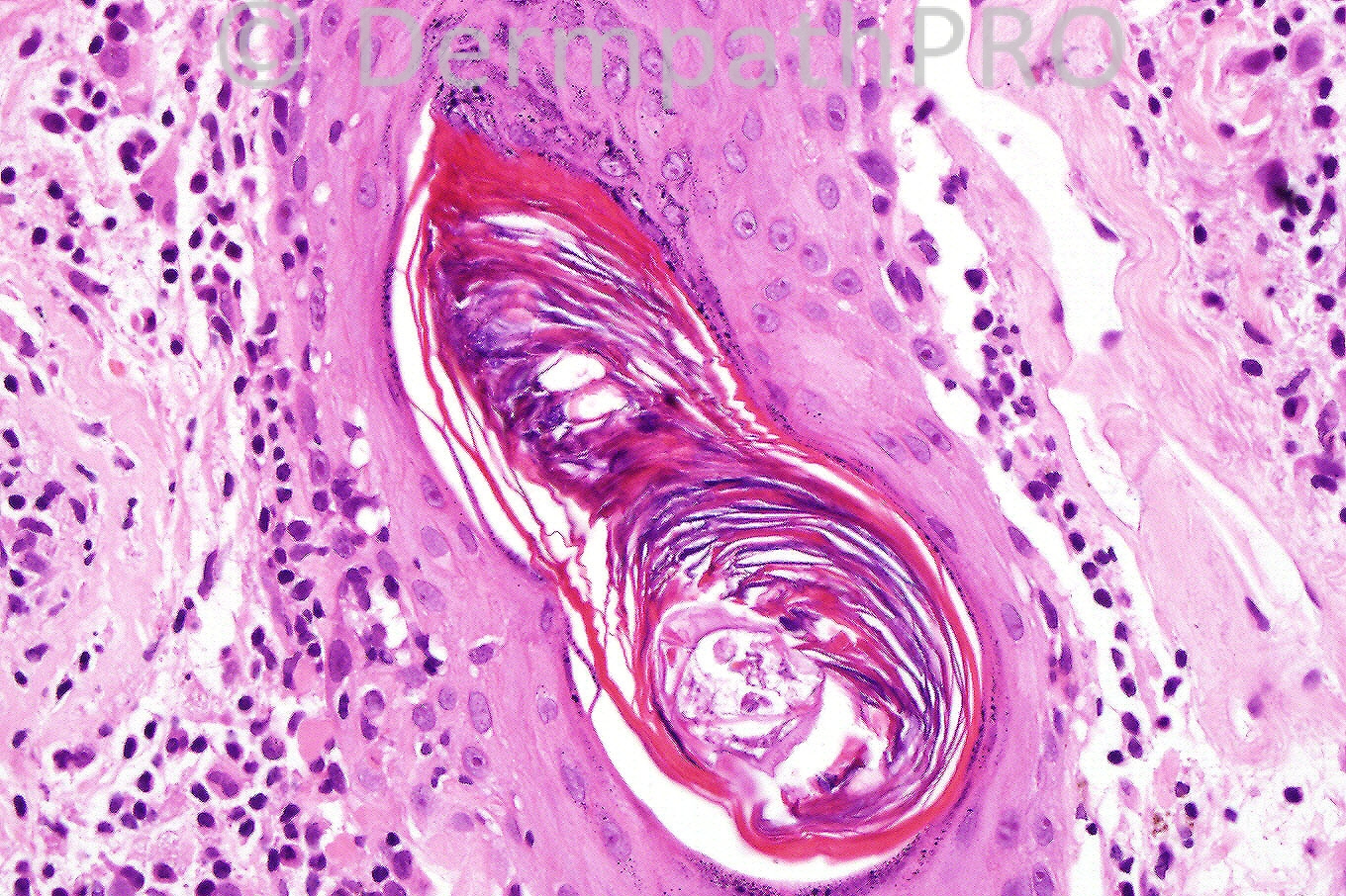
Female 34 years with scaly erythematous plaques on her cheeks.


  Report Record

User Feedback


[b][color=#000000]Lupus erythematosus[/color][/b].

Share this comment


Link to comment
share_externally

Guest Francisco Vílchez-Márquez

Posted

Discoid lupus

Share this comment


Link to comment
share_externally

Guest Hazem Hamed

Posted

Discoid lupus erythematosus.

Share this comment


Link to comment
share_externally

Guest Amr Gohar

Posted

A lichenoid reaction pattern and a superficial and deep dermal infiltrate of inflammatory cells with a tendency to accumulate around the pilosebaceous
follicles. Keratotic follicular plugging is among other features. Discoid lupus erythematosus (DLE).

Share this comment


Link to comment
share_externally

Sasi Attili

Posted

discoid lupus

Share this comment


Link to comment
share_externally

Admin_Dermpath

Posted

A beautiful case of [b]Lupus. [/b]

I would recommend, if possible, users set up an account (it is free and does not require you to subscribe) so you can easily sign in when you make a comment! Also your profile will allow others to know a little bit more about you and if needed they can send an internal communication.

Regards


Dr Iskander H. Chaudhry
Consultant Dermatopathologist
Adult Histopathology
Central Manchester University Hospitals NHS Foundation Trust
Hospital
Oxford Road, Manchester M13 9WL

Share this comment


Link to comment
share_externally

Guest Dr Engin Sezer

Posted

Tortuous thickening of the basal membrane with follicular plug together with superficial and deep perivascular and periadnexal lymphocytic infiltration consistent with DLE, beautiful images.

Share this comment


Link to comment
share_externally

Dr. Richard Carr

Posted

I have added a blog on Friday's case of regressing KA. Regards to all.

Share this comment


Link to comment
share_externally

[font=courier new,courier,monospace]Thanks Richard.[/font]

Share this comment


Link to comment
share_externally

Dr. Phillip McKee

Posted

DLE is correct. As I remember, it is a case that Richard very kindly shared with me. One of the great things about the dermatopathology community is the willingness to share material. I have so many people from all over the world who regularly mail me either difficult cases or beautiful examples. It reflects that at heart, we are all just people and that there really are no borders!

Share this comment


Link to comment
share_externally

Dr. Phillip McKee

Posted

I also meant to add support to Iskander's comment. It will cost you nothing to sign in and be able to comment on the spot diagnoses. You will also be able to have a look at the rest of the site and see if it is for you. It is a very active website that changes daily and a wonderful way to establish a truly global community of like-minded people.

Share this comment


Link to comment
share_externally


×
×
  • Create New...