Jump to content
In this section we have spot diagnoses posted on a daily basis since June 2010, now over 4000! You can review the archived cases and read the suggested diagnoses by users and the final comment by the contributors.
Case are uploaded each week day by 10 am UK time with the correct diagnosis will generally be posted at 8 pm UK time. Why not view the most recent spot diagnosis and proffer a diagnosis?

Case Number : Case 547 - 13 July Posted By: Guest

Please read the clinical history and view the images by clicking on them before you proffer your diagnosis.
Submitted Date :
   (0 reviews)

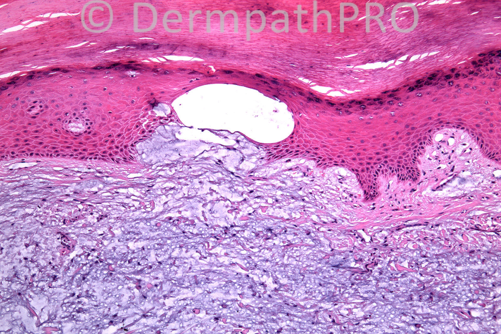
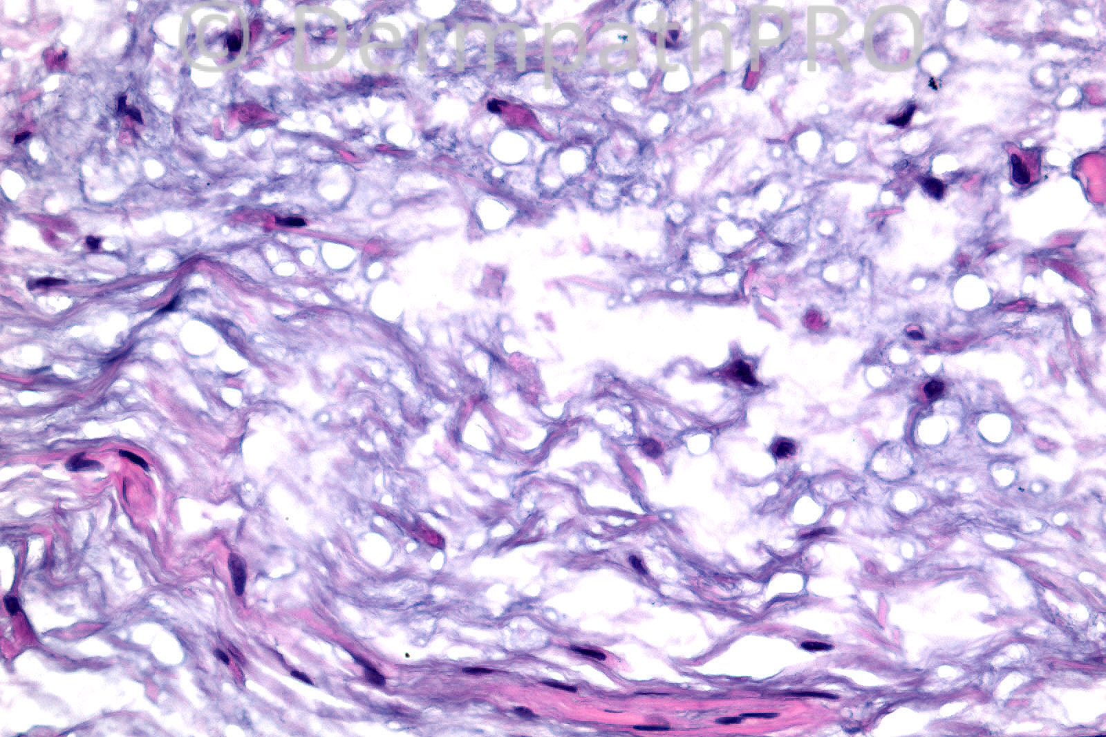
Male 56 years, ?Callus between webspace of thumb and forefinger.

Case posted by Dr. Richard Carr


  Report Record

User Feedback


Guest Dr. Francisco Vílchez

Posted

Focal mucinosis

Share this comment


Link to comment
share_externally

Guest Hazem Hamed

Posted

[b]Digital Mucous/myxoid cyst.[/b]

Share this comment


Link to comment
share_externally

[b]Digital myxoma (a.k.a. digital mucus cyst).[/b]

Share this comment


Link to comment
share_externally

Guest Hazem Hamed

Posted

[b]I would like to add myxoid neurofibroma and myxoid neurothekoma in the DD (S100 and EMA would help).[/b]

Share this comment


Link to comment
share_externally

[size=4][font=courier new,courier,monospace][color=#231F20][color=#231F20]Myxomatous change. Considering the clinical data too, I would suggest [/color][color=#000000][b]superficial acral fibromyxoma[/b] or [/color][color=#231F20][b]m[b]yxoid [/b]cyst,[/b] possibly [b]traumatic[/b] in this site. [/color][/color][/font][/size]

Share this comment


Link to comment
share_externally

Guest Juan Carlos Garcés, Ecuador

Posted

Digital mixoid cyst

Share this comment


Link to comment
share_externally

Sasi Attili

Posted

To me, its a bit too cellular to be a simple myxoid cyst. The spindle and stellate cells made me favour a superficial acral fibromyxoma.

However in the images on the bottom (esp the bottom right), there are a few cells which do look pleomorphic and atypical. Thus I do wonder about acral myxoinflammatory fibroblastic sarcoma. But atypical cells have been described in Superficial acral angiomyxoma and I am not really sure how to make the distinction.

Share this comment


Link to comment
share_externally

[size=4][font=courier new,courier,monospace]Well, my[color=#231F20]xoinflammatory fibroblastic sarcoma (many cases are clinically diagnosed as ganglion cysts), shows [/color][color=#000000][b]numerous inflammatory cells[/b] in addition to the [/color][color=#231F20][b]neoplastic[/b] [b]cells[/b]. [b]Immunohistochemistry[/b] is useful too. [/color][/font][/size]

Share this comment


Link to comment
share_externally

Dr. Richard Carr

Posted

Well this in fact came with the additional history that he was tree surgeon
and the area was being repeatedly traumatised during his work. Consequently
our diagnosis was a mucus cyst (traumatic type). I have seen very few
examples of traumatic type mucus cyst as by far the more common type of
digital mucus cyst we see is the ganglionic type usually presenting overlying a distal
interphalyngeal joint. In the latter you more often than not can see a
deeper component lined by synoviocytes. I agree without the context one could
consider the differential diagnoses of superficial acral fibromyxoma and
angiomyxoma. You probably already know I think soft tissue pathology is a dark
art!

Share this comment


Link to comment
share_externally


×
×
  • Create New...