Jump to content
In this section we have spot diagnoses posted on a daily basis since June 2010, now over 4000! You can review the archived cases and read the suggested diagnoses by users and the final comment by the contributors.
Case are uploaded each week day by 10 am UK time with the correct diagnosis will generally be posted at 8 pm UK time. Why not view the most recent spot diagnosis and proffer a diagnosis?

Case Number : Case 548 - 16 July Posted By: Guest

Please read the clinical history and view the images by clicking on them before you proffer your diagnosis.
Submitted Date :
   (0 reviews)

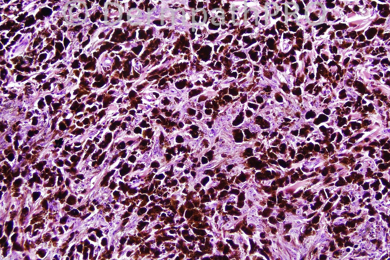
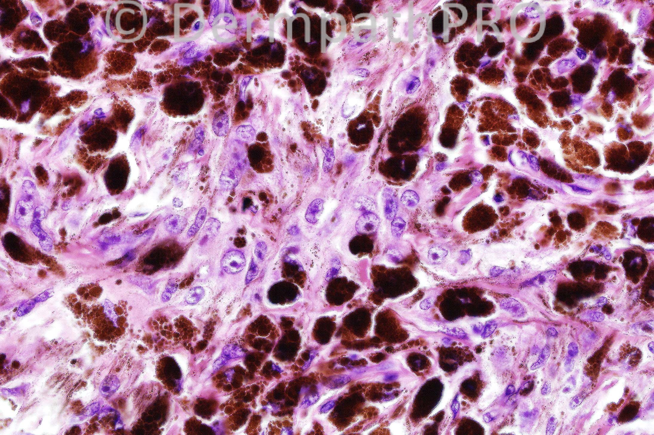
Male 42 years.  Pigmented nodule on face.


  Report Record

User Feedback


Guest Hazem Hamed

Posted

[b]My diagnosis is Pigmented Epithelioid melanocytoma (no mitosis, ulceration or junctional component). DD animal type melanoma. [/b]

Share this comment


Link to comment
share_externally

Epithelioid blue naevus.

Share this comment


Link to comment
share_externally

Guest Hazem Hamed

Posted

[b]Also epithelioid blue nevus is in the differntial diagnosis.[/b]

Share this comment


Link to comment
share_externally

Sasi Attili

Posted

My preferred diagnosis based on lack of infiltrative deep margins, good circumscription/ symmetry and lack of significant atypia/ mitoses, is Epithelioid Blue nevus.

However I think this would fall into the controversial pigmented epithelioid melanocytoma category which encompasses Epithelioid Blue nevus and Animal-type melanoma!

Share this comment


Link to comment
share_externally

Blue nevus vs pigmented epitheloid melanocytoma

Share this comment


Link to comment
share_externally

Guest Juan Carlos Garcés, Ecuador

Posted

Pigmented Epithelioid melanocytoma

Share this comment


Link to comment
share_externally

Guest MarieMD

Posted

Epithelioid blue nevus

Share this comment


Link to comment
share_externally

Guest Sadiq M Amer

Posted

Epithelioid blue nevus.

Share this comment


Link to comment
share_externally

Dr. Phillip McKee

Posted

I think that it is not possible to make a distinction between epithelioid blue nevus and pigment synthesizing melanoma on these images. They could represent either. I believe however that there were mitoses in the original section but the slide that I photographed was a recut devoid of mitotic activity. The case was diagnoses as a pigment synthesizing melanoma. I have no follow-up information.

Share this comment


Link to comment
share_externally

[size=4][font=arial,helvetica,sans-serif][color=#231F20]The blue naevus is characterized clinically by its colour (Tyndall effect, [color=#000000]the shorter wavelength of light [blue] is scattered/reflected by the deep brown pigment[/color]) and symmetrical shape and must be differentiated from other dermal melanoses. [/color][color=black]The term ‘pigmented epithelioid melanocytoma’ is used by some for a ‘low-grade melanocytic tumour with metastatic potential indistinguishable from animal-type [equine - resembling the usually benign tumours seen in many grey horses] melanoma and epithelioid blue naevus’. When in doubt the lesion is managed as for melanoma. [/color][/font][/size]

Share this comment


Link to comment
share_externally

[color=#000000][url="http://www.ncbi.nlm.nih.gov/pubmed?term=Antony%20et%20al%20%20%E2%80%98pigment%20synthesizing%20melanoma"]Antony et al[/url] in the United Kingdom have suggested ‘pigment synthesizing melanoma’ as a preferable term to ‘pigmented epithelioid melanocytoma’.[/color]

Share this comment


Link to comment
share_externally


×
×
  • Create New...