Jump to content
In this section we have spot diagnoses posted on a daily basis since June 2010, now over 4000! You can review the archived cases and read the suggested diagnoses by users and the final comment by the contributors.
Case are uploaded each week day by 10 am UK time with the correct diagnosis will generally be posted at 8 pm UK time. Why not view the most recent spot diagnosis and proffer a diagnosis?

Case Number : Case 549 - 17 July Posted By: Guest

Please read the clinical history and view the images by clicking on them before you proffer your diagnosis.
Submitted Date :
   (0 reviews)

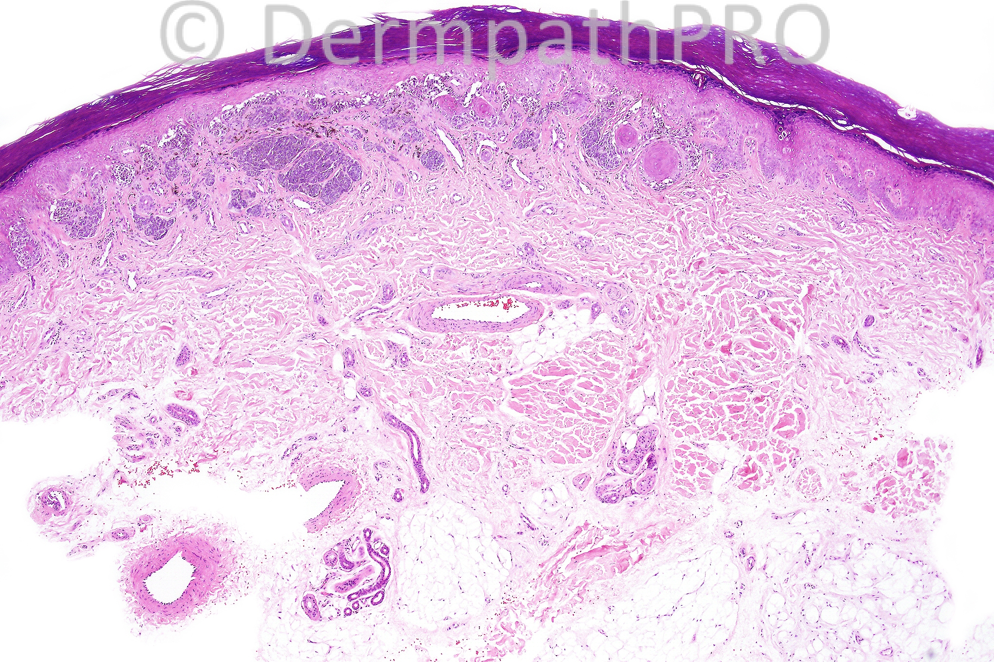
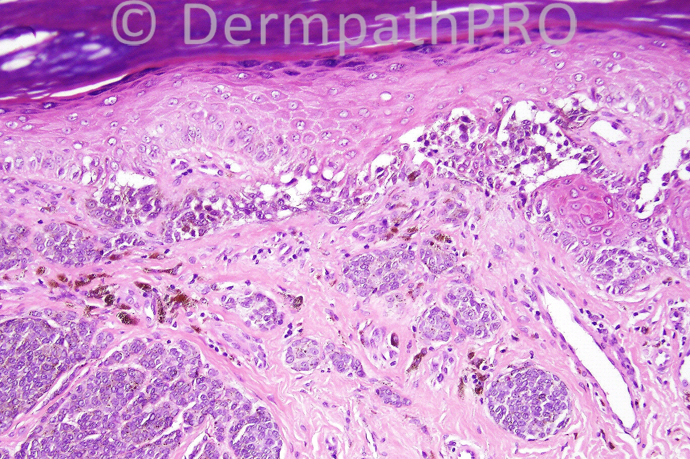
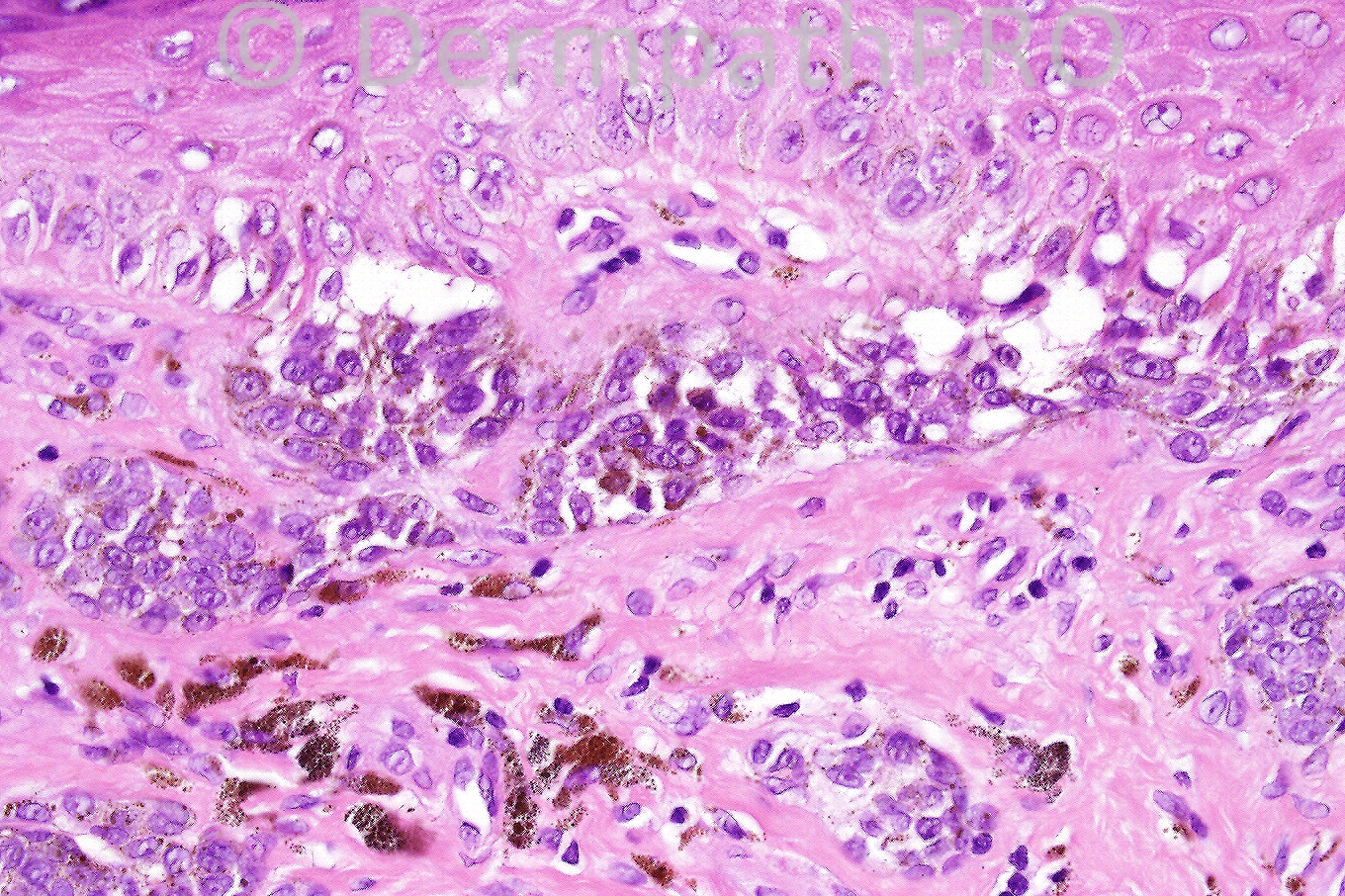
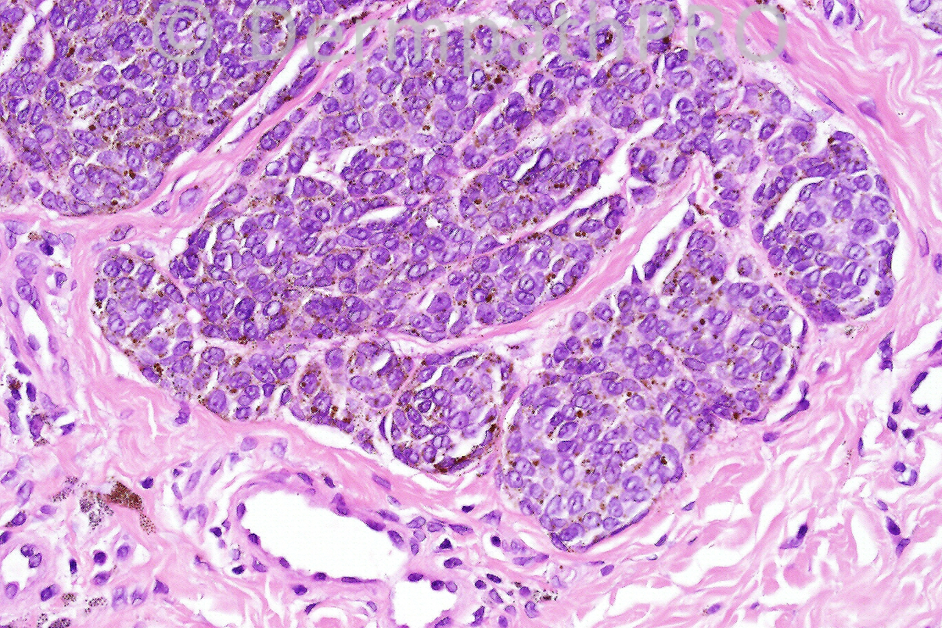
Male 40 years, pigmented lesion on foot.

We are grateful to Dr. Richard Carr for this case.


  Report Record

User Feedback


Sasi Attili

Posted

Compound acral nevus with site specific features

Share this comment


Link to comment
share_externally

Guest Dr. Francisco Vílchez

Posted

Acral nevus.

Share this comment


Link to comment
share_externally

Acral compound nevus

Share this comment


Link to comment
share_externally

Guest Dr Engin Sezer

Posted

Compound nevus on specific site

Share this comment


Link to comment
share_externally

Guest Juan Carlos Garcés, Ecuador

Posted

Acral compound nevus

Share this comment


Link to comment
share_externally

Guest MarieMD

Posted

compound nevus on acral skin

Share this comment


Link to comment
share_externally

Guest Hazem Hamed

Posted

[font=tahoma, geneva, sans-serif]I agree with Compound nevus on specific site but my only concern is nevoid melanoma (though there is no mitosis). I can not see evidence of maturation[/font].

Share this comment


Link to comment
share_externally

Compound naevus. Both the junctional and superficial dermal components are shown.

Share this comment


Link to comment
share_externally

Victor Delgado

Posted

Acral compound nevus

Share this comment


Link to comment
share_externally

Guest Ixtabay, Yucatán

Posted

Acral compound nevus

Share this comment


Link to comment
share_externally

Dr. Richard Carr

Posted

Yes this was a case of a benign acral melanocytic naevus.
Apologies for the delay in posting the diagnosis.

Share this comment


Link to comment
share_externally


×
×
  • Create New...