Jump to content
In this section we have spot diagnoses posted on a daily basis since June 2010, now over 4000! You can review the archived cases and read the suggested diagnoses by users and the final comment by the contributors.
Case are uploaded each week day by 10 am UK time with the correct diagnosis will generally be posted at 8 pm UK time. Why not view the most recent spot diagnosis and proffer a diagnosis?

Case Number : Case 553 - 23 July Posted By: Guest

Please read the clinical history and view the images by clicking on them before you proffer your diagnosis.
Submitted Date :
   (0 reviews)

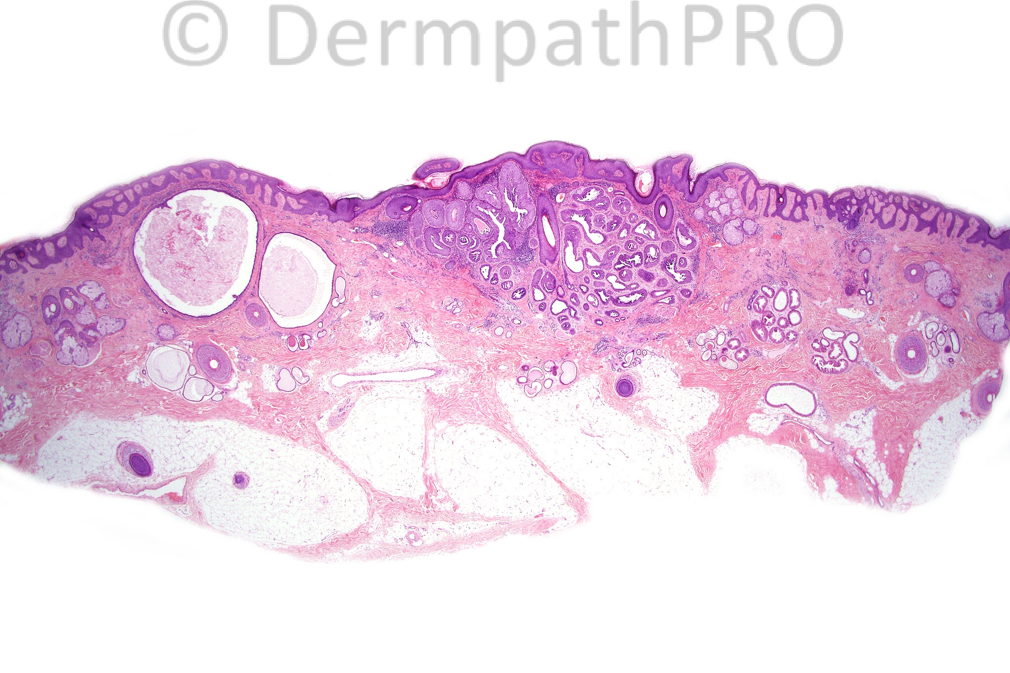
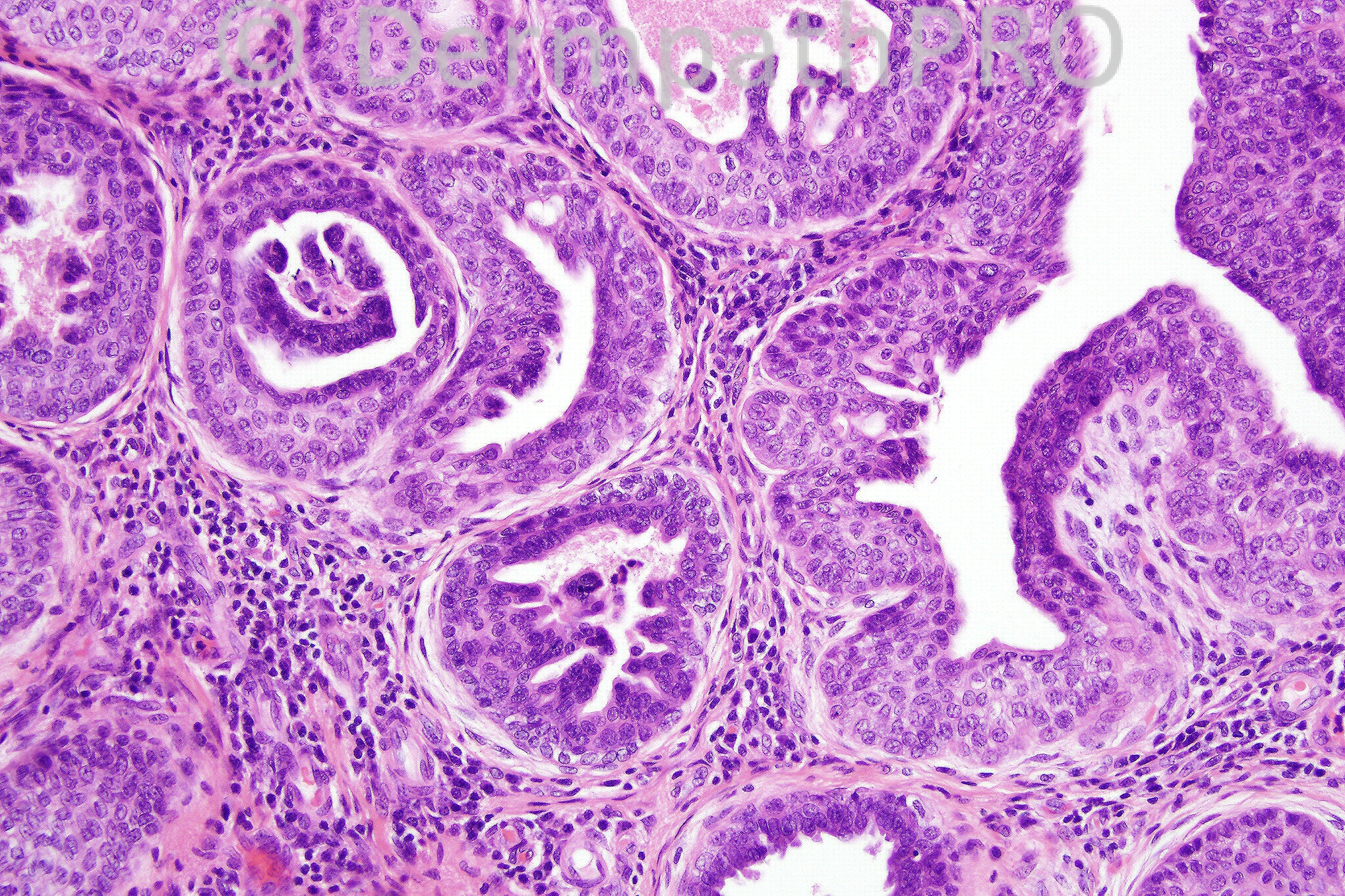
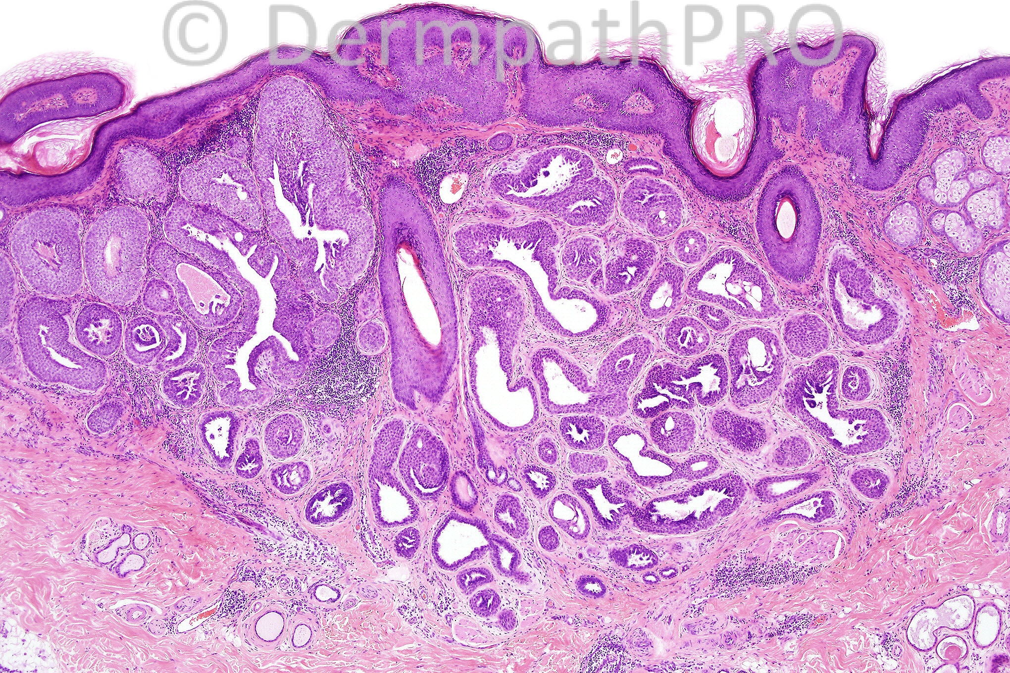
Female 40 year old, lesion on scalp.

  Report Record

User Feedback


Guest Hazem Hamed

Posted

[b]Papillary eccrine adenoma arising in a background of organoid nevus. Though few inflammatory cells including occasional plasma cells are present the features are not typical of syringocystadenoma papilleferum. [/b]

Share this comment


Link to comment
share_externally

Guest Dr. Francisco Vílchez

Posted

Papillary projections, no decapitation, and typical location: agree, papillary eccrine adenoma.

Share this comment


Link to comment
share_externally

Adenoma, be it eccrine or apocrine, tubular or papillary.

Share this comment


Link to comment
share_externally

Guest Dr Engin Sezer

Posted

[i][b]Duct-like structures in the deeper layer [/b][/i]together with apocrine epithelial elements and plasma cells consistent with [b]syringocystadenoma papilliferum[/b].

Share this comment


Link to comment
share_externally

nevus sebaceous with a tubular apocrine adenoma

Share this comment


Link to comment
share_externally

Guest amira tawdy

Posted

Papillary eccrine adenoma

Share this comment


Link to comment
share_externally

Guest MarieMD

Posted

[b][font="Arial"][size="2"][color="#000080"]Tubulopapillary hidradenoma (PEA) arising in Nevus Sebaceous[/color][/size][/font][/b]

Share this comment


Link to comment
share_externally

Guest Juan Carlos Garcés, Ecuador

Posted

papillary ecrine adenoma in nevus sebaceous

Share this comment


Link to comment
share_externally

Sasi Attili

Posted

Cystadenoma + papillary eccrine adenoma arising in an organoid nevus.

Share this comment


Link to comment
share_externally

Dr. Mona Abdel-Halim

Posted

I favor tubular apocrine adenoma (tubulopapillary hidradenoma) on top of organoid nevus

Share this comment


Link to comment
share_externally

[color=#231F20][b]Tubular apocrine adenoma[/b]. As with many adnexal tumours, [b]histopathological[/b] [b]overlap between tubular apocrine adenoma and papillary eccrine adenoma [/b]is not uncommon. Overlapping features with syringocystadenoma papilliferum are also seen, however, in classic [/color][color=#000000]syringocystadenoma[/color][color=#231F20] [/color][color=#000000]papilliferum, the inflammatory cells, particularly plasma cells, should be more[/color][color=#231F20] [/color][color=#000000]prominent. Apocrine cystadenomas have true papillae with a fibrous core.[/color]

Share this comment


Link to comment
share_externally

Dr. Phillip McKee

Posted

I favored a diagnosis of tubular apocrine adenoma arising in an organoid nevus. Obviously I am well aware of the debate between apocrine or eccrine. It seems to me to be a rather futile exercise but being old fashioned, I restrict the term ecrrine tubular adenoma to those on the distal limbs. Although not very convincing, there is just a hint of aporcine secretion in the last image or perhaps it is just fanciful thinking.
On another matter, I have made two posts on my page of facebook today. I would really appreciate it if you could go to those comments and post your thoughts on either or both posts, preferably both. It would be very helpful to Iskander and myself and very much appreciated.

Share this comment


Link to comment
share_externally


×
×
  • Create New...