Jump to content
In this section we have spot diagnoses posted on a daily basis since June 2010, now over 4000! You can review the archived cases and read the suggested diagnoses by users and the final comment by the contributors.
Case are uploaded each week day by 10 am UK time with the correct diagnosis will generally be posted at 8 pm UK time. Why not view the most recent spot diagnosis and proffer a diagnosis?

Case Number : Case 557 - 27 July Posted By: Guest

Please read the clinical history and view the images by clicking on them before you proffer your diagnosis.
Submitted Date :
   (0 reviews)

Male 54 years, back, 6/12 history of rash trunk and proximal limbs, comes and goes, multiple papules and plaques ?urticaria.

Case posted by Dr. Richard Carr


  Report Record

User Feedback


Guest Hazem Hamed

Posted

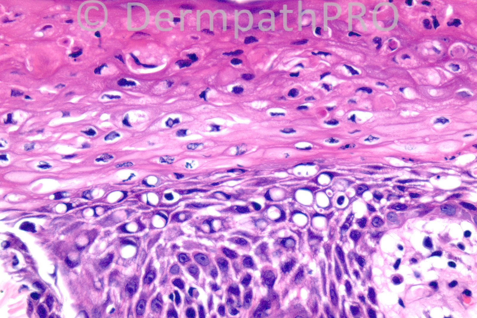
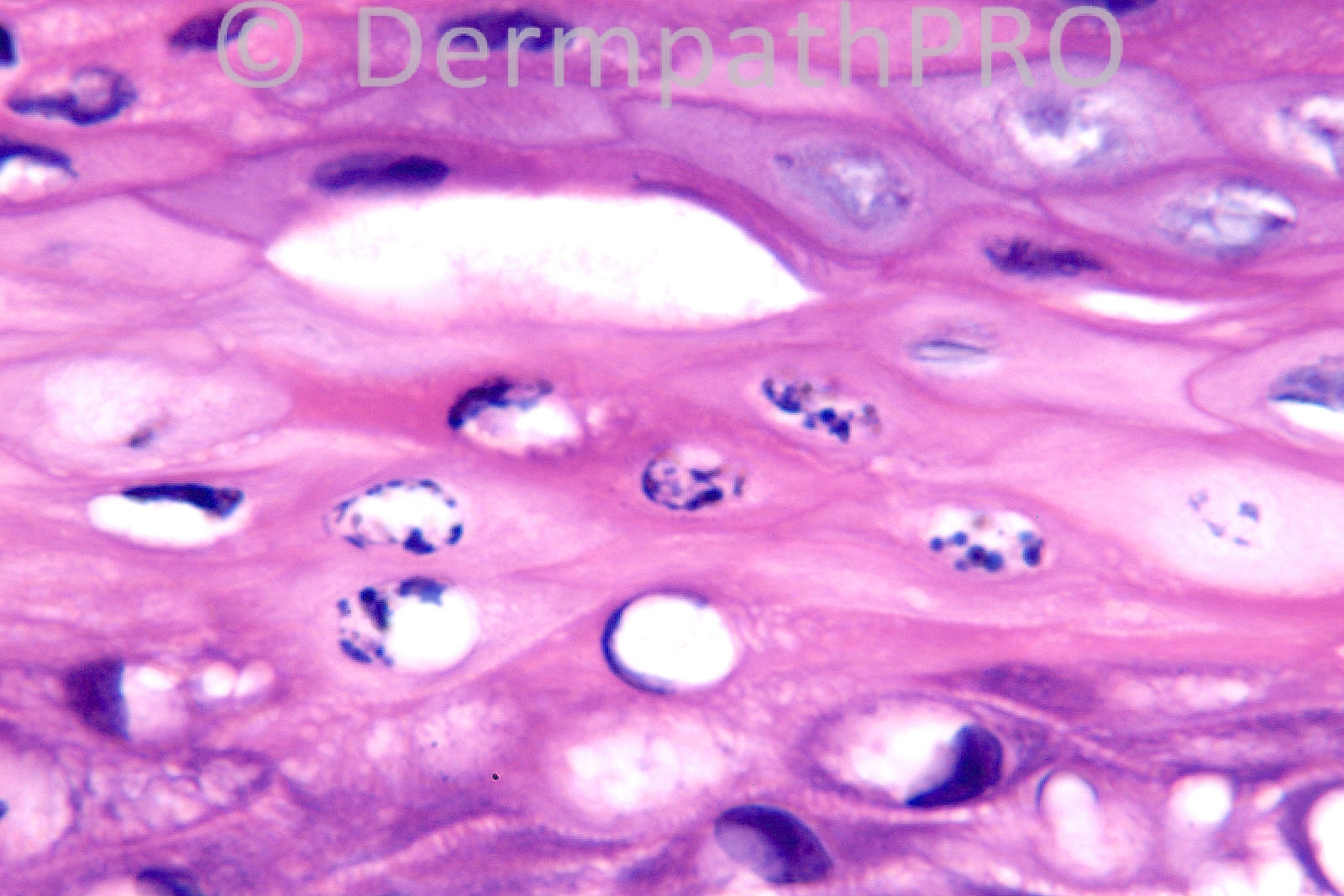
[b]Necrolytic migratory erythema (glucagonoma syndrome) is my first consideration. Other diseases with similar histological findings include Pellagra and zinc deficiency.[/b]

Share this comment


Link to comment
share_externally

Necrolytic erythema.

Share this comment


Link to comment
share_externally

The histopatological findings suggest any of the nutritional deficiencies (necrolytic migratory erythema or zinc deficiency -acrodermatitis enteropathica), however, the clinical information does not support the diagnosis.

Share this comment


Link to comment
share_externally

Sasi Attili

Posted

Completely agree with Marcela.

Share this comment


Link to comment
share_externally

Guest Dr Gonzalo de Toro

Posted

I agree with Marcela too. Maybe could be a Glucagonoma syndrome with psoriasiform hyperplasia

Share this comment


Link to comment
share_externally

Guest MarieMD

Posted

necrolytic migratory erythema secondary to either glucagonoma or pseudoglucagonoma syndrome. (Histology is that of nutritional deficiency)

Share this comment


Link to comment
share_externally

[font=arial,helvetica,sans-serif][size=4][color=black][b]Vacuolated keratinocytes with scattered dyskeratotic cells in the upper epidermis[/b],[b] necrotic keratinocytes [/b]are associated with clefts and the term[/color][color=#000000] [color=black]‘necrolysis’ is used. [b]Parakeratosis with a degree of acanthosis[/b]. [/color][b]Mo[b]derate [/b]perivascular infiltrate of lymphoid cells [/b]in the dermis. [b]Necrolytic migratory erythema [/b](waves of extending annular or circinate itchy erythema with [/color][color=#231F20]a prolonged, fluctuating course [[/color][color=#000000]resolution of involved areas usually takes place within two weeks]). The skin lesions are polymorphous but the primary lesions are erythematous patches/plaques. As always, clinicopathological correlation is vital. [color=#231F20]Necrolytic migratory erythema is a cutaneous reaction that needs to be carefully investigated. Among the investigations are determination of glucose, zinc and plasma glucagon levels and pancreatic/hepatic scans. [/color][/color][/size][/font]

Share this comment


Link to comment
share_externally

necrolytic migratory erythema

Share this comment


Link to comment
share_externally

Dr. Mona Abdel-Halim

Posted

Necrolytic migratory erythema

Share this comment


Link to comment
share_externally

Dr. Richard Carr

Posted

Well I think everyone is in aggreement with the possible histological differenatial diagnoses in this case (i.e. necrolytic migratory erythema and nutritional deficiencies). Certainly the histology is very striking! I offered exactly the same possibilities as those listed above. Unfortunately for reasons I cannot divulge further investigation was not possible and frustratingly we do not know the final aetiology in this case. Thank you for your helpful comments and discussion and regards to all.

Share this comment


Link to comment
share_externally

Thank you Richard.

Share this comment


Link to comment
share_externally


×
×
  • Create New...