Jump to content
In this section we have spot diagnoses posted on a daily basis since June 2010, now over 4000! You can review the archived cases and read the suggested diagnoses by users and the final comment by the contributors.
Case are uploaded each week day by 10 am UK time with the correct diagnosis will generally be posted at 8 pm UK time. Why not view the most recent spot diagnosis and proffer a diagnosis?

Case Number : Case 558 - 30 July Posted By: Guest

Please read the clinical history and view the images by clicking on them before you proffer your diagnosis.
Submitted Date :
   (0 reviews)

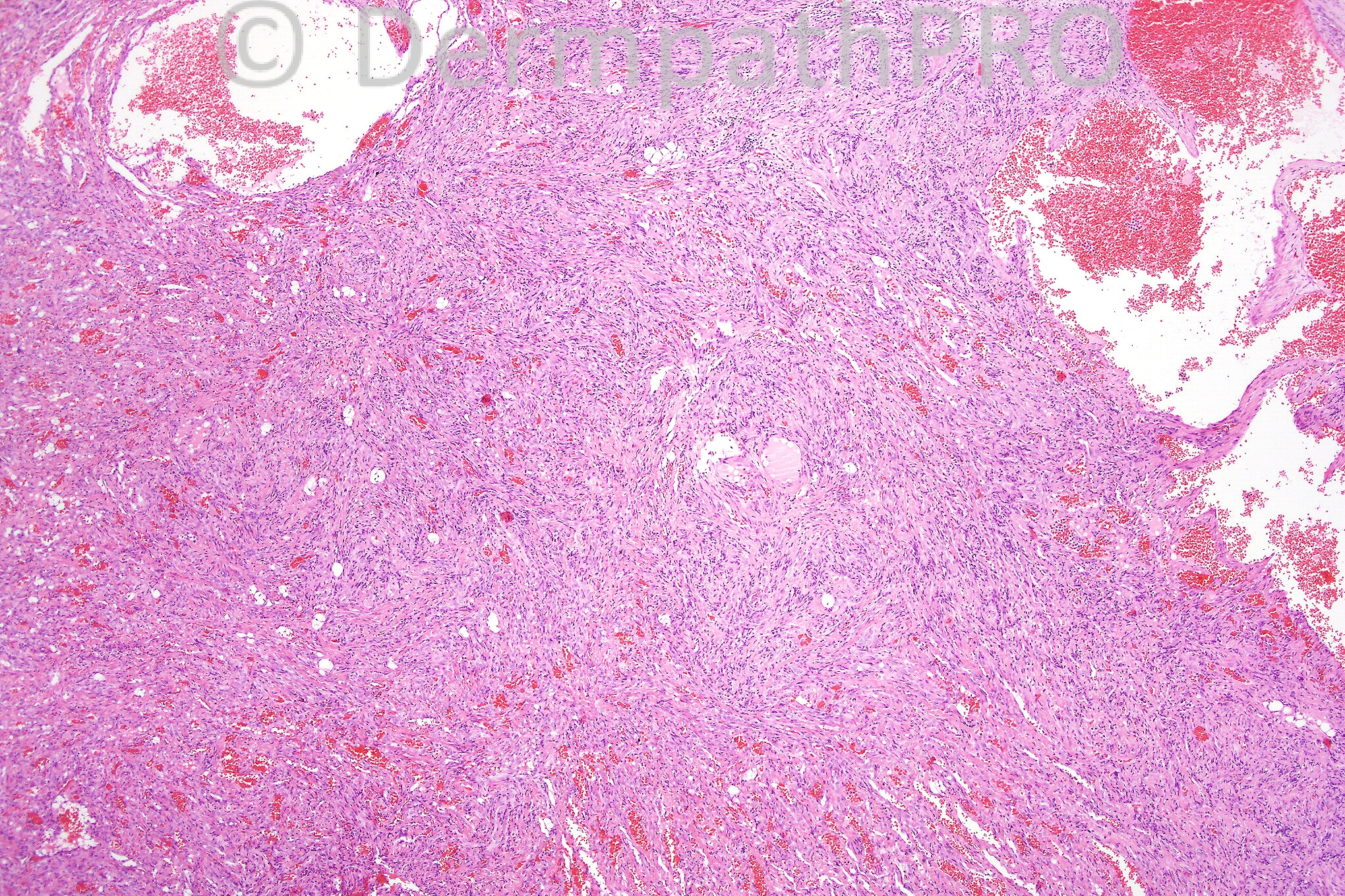
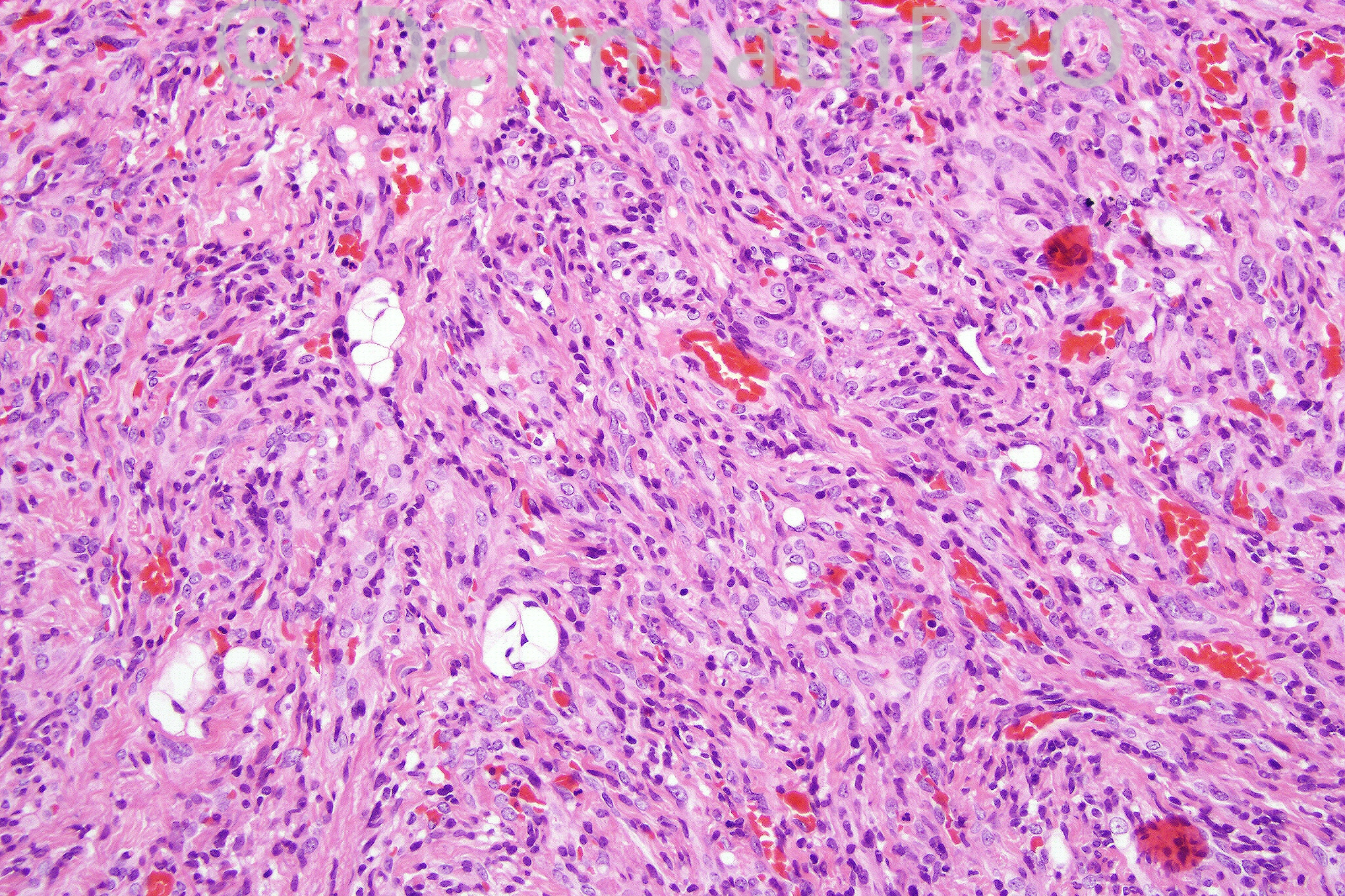
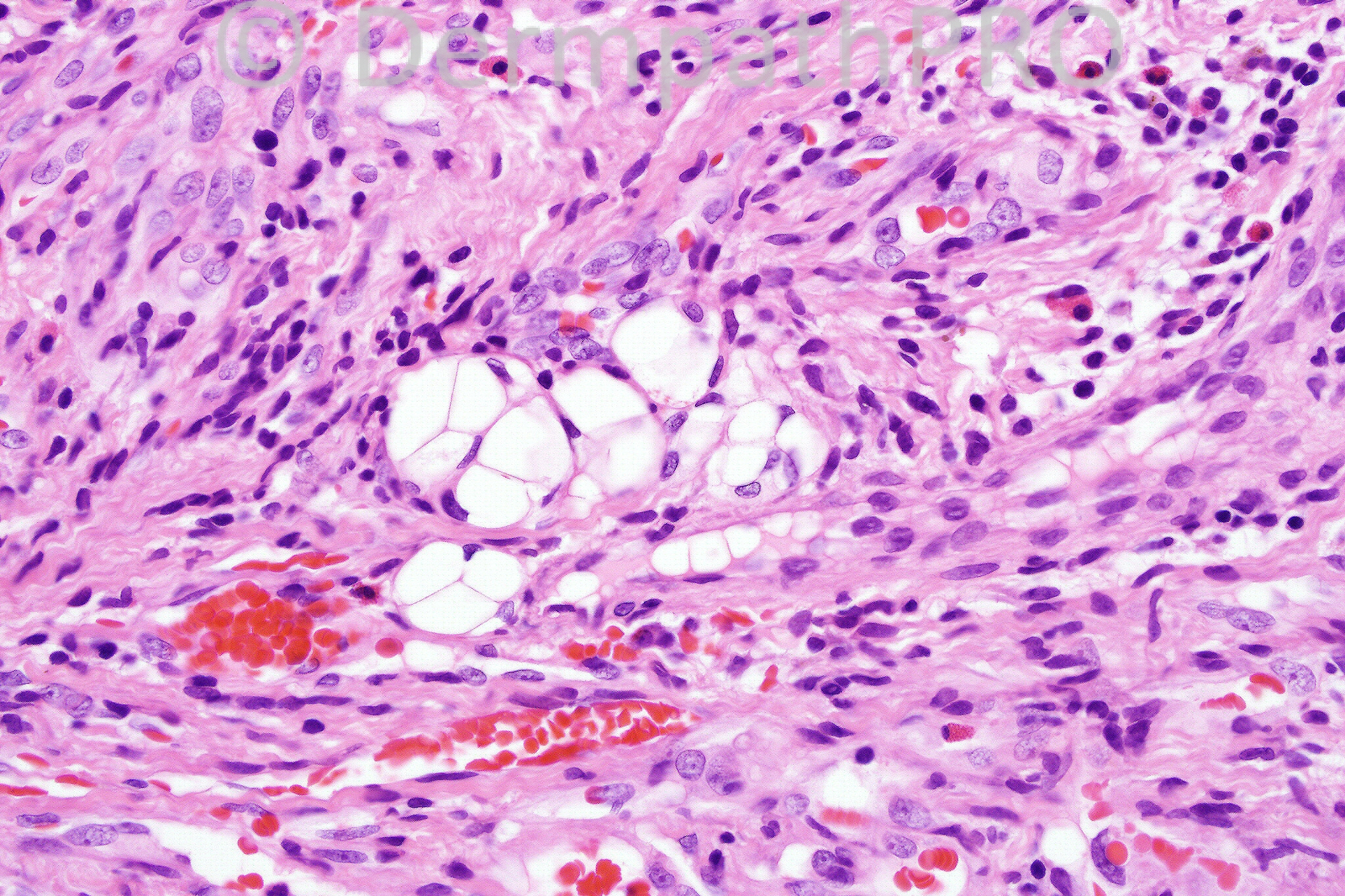
Age and sex unknown. Nodule on finger.

We are grateful to Dr. Richard Carr who has provided this case.


  Report Record

User Feedback


Guest Hazem Hamed

Posted

Thought of spindle cell/kaposiform haemangioendothelioma. Intracytoplasmic lumina are present in the endothelial cells in the 2nd image.The multivacuolated cells are either pseudolipoblasts or multi vaculated endothelial cells !!. HHV8 is required to exclude kaposi sarcoma.

Share this comment


Link to comment
share_externally

Guest Dr. Francisco Vílchez

Posted

Vascular channels. Without clinical information is difficullt, though i´m thinking in Kaposi Sarcoma. Spindle cell hemangioendothelioma is a good differential diagnosis.

Share this comment


Link to comment
share_externally

Guest Dr Gonzalo de Toro

Posted

spindle cell hemangioma/haemangioendothelioma. HHV8 is necessary to rule out kaposi sarcoma

Share this comment


Link to comment
share_externally

[color=#000000]Provisionally, a [/color][b][color=#231F20]spindle cell haemangioma. [/color][/b][color=#231F20]T[/color][color=#000000]hin-walled cavernous-like [b]c[b]ongested channels[/b] –[/b] [b]slit like spaces[/b] - [b]spindle cells[/b] – [b]“[/b][/color][b][color=#231F20]epithelioid” cells (lying in groups) with prominent vacuolation[/color][/b][color=#000000]. [/color][color=#231F20]Immunohistochemistry to [/color][color=#000000]check the endothelial nature of the cells and any [/color][color=#231F20]focal staining for actin. Also, testing for [/color][color=#000000]HHV-8. [/color]

Share this comment


Link to comment
share_externally

Guest Juan Carlos Garcés, Ecuador

Posted

spindle cell hemangioma

Share this comment


Link to comment
share_externally

Guest MarieMD

Posted

spindle cell hemangioma/haemangioendothelioma

Share this comment


Link to comment
share_externally

Dr. Mona Abdel-Halim

Posted

Spindle cell hemangioma (hemangioendothelioma) vs. Kaposiform hemangioendothelioma. Would love to read a way of differentiating between them.

Share this comment


Link to comment
share_externally

KS vs spindle cell hemangioendothelioma

Share this comment


Link to comment
share_externally

Dr. Phillip McKee

Posted

Difficult case but the presence of so many endothelial cell lumina is very much in favor of spindle cell hemangioma. I don't know whether HHV-8 was done but assume that it was negative. Kaposiform hemangioendothelioma is another good differential diagnosis. My personal experience of this tumor is limited but msot importantly it is HHV-8 negative, often shows mixed growth pattern- KS-like areas, foci more in keeping with capillary hemangioma and sometimes areas resembling tufted hemagioma and even lymphangioma. It is commonly associated with the Kasabach-Merritt syndrome.

Thanks to all of you who have subscribed to the site so far. Have you given thought to an institutional subscription? This is such good value- a whole year for up to 15 members of staff. You can access the site anywhere you are- at work, at home or even at the airport!! Folks in training will get such a wealth of experience. It really is a must-have!!

Share this comment


Link to comment
share_externally

Dr. Mona Abdel-Halim

Posted

Thanks Dr McKee :-)

Share this comment


Link to comment
share_externally


×
×
  • Create New...