Jump to content
In this section we have spot diagnoses posted on a daily basis since June 2010, now over 4000! You can review the archived cases and read the suggested diagnoses by users and the final comment by the contributors.
Case are uploaded each week day by 10 am UK time with the correct diagnosis will generally be posted at 8 pm UK time. Why not view the most recent spot diagnosis and proffer a diagnosis?

Case Number : Case 517 Posted By: Guest

Please read the clinical history and view the images by clicking on them before you proffer your diagnosis.
Submitted Date :
   (0 reviews)

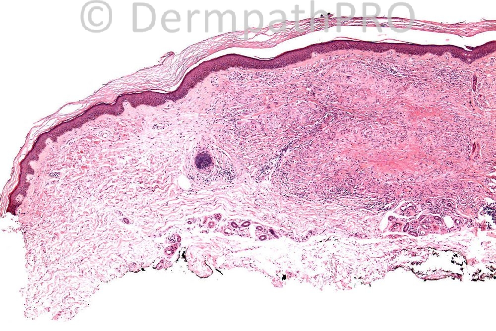
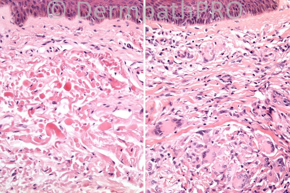
Male 70 years, annular lesion with atrophic centre on hand: dorsum..


  Report Record

User Feedback


Admin_Dermpath

Posted

[b]Geoff O'Brien - Healthscope Pathology (Sydney) Wrote:[/b]

I favour actinic granuloma also. Thank you Dr Carr for the nice exposition. My late father accepted some necrobiosis as appears to be present here. Interestingly his 1975 paper mentioned a case with actinic granuloma of the neck, and a lesion on the hand with mixed AG and GA features. He felt Hanke's 1979 paper proposing AEGCG may have ignored the subtle and pervasive presence of actinic elastosis. In contrast with GA, with AG we can offer the patient a causation -solar and other radiant exposure.

[size=2]Submitted on 29/06/2012 06:43[/size]
[b]Richard Carr - Warwick (UK) Wrote:[/b]

Apologies for the delay but you are all on track. In fact I had posted 4 images originally but only 2 made it on to the website. We did do an elastic stain and there were indeed three zones to this lesion a large central area with no elastic at all and subtle dermal fibrosis (more extensive than typical GA). An intermediate zone of active giant cell reaction and elastophagocytosis and an outer area with prominent intact orange elastic fibre. We diagnosed this case as in keeping with the clinical diagnosis if actinic granuloma (I ommited the additional history of excessive solar elastosis on the face). By the way whenever you see giant cells or histiocytes in any reaction in the skin you will inevitably see elastophagocytosis as a non-specific phenomenon so it is more the overrall features here that make a cinicopathological diagnosis not elastophagocytosis per se. I will leave it to you all to decide if you think actinic granuloma (O'Brien granuloma) is a variant of GA as some very eminent dermatopathologists opine). However usually GA is patchy dermal with areas of intact normal dermis (not the case in this particular case but that is difficult from the images you were given). It emphasises the need for a good incision biopsy across the edge to include the centre of the lesion.

[size=2]Submitted on 10/06/2012 11:39[/size]
[b]Engin Sezer - (Istanbul) Wrote:[/b]

I've never seen Dr. Marie with an incorrect diagnosis and I've now realized that annular elastolytic giant cell granuloma is in the spectrum of elastolytic granulomata (such as actinic granuloma (O'Brien), which may also be observed in the absence of solar elastosis, hence annular elastolytic giant cell granuloma!

[size=2]Submitted on 05/06/2012 15:21[/size]
[b]rabia - shifa (karachi) Wrote:[/b]

granuloma annulare

[size=2]Submitted on 05/06/2012 15:03[/size]
[b]rabia - shifa (karachi) Wrote:[/b]

granuloma annulare

[size=2]Submitted on 05/06/2012 15:03[/size]
[b]Marie MD - () Wrote:[/b]

annular elastolytic giant cell granuloma--

[size=2]Submitted on 04/06/2012 16:44[/size]
[b]Eman El-Nabarawy - () Wrote:[/b]

Actinic granuloma.

[size=2]Submitted on 04/06/2012 16:02[/size]
[b]Rodrigo Restrepo - UPB/CES (Mdlln/Col) Wrote:[/b]

Granuloma annulare

[size=2]Submitted on 04/06/2012 12:55[/size]
[b]Yüksel Okumuş - Bursa State Hospital (Turkey) Wrote:[/b]

Granuloma annulare or necrobiosis lipoidica

[size=2]Submitted on 04/06/2012 12:53[/size]
[b]Marcela Saeb Lima - INSMNSZ (Mexico City) Wrote:[/b]

GA, though the clinical history suggests an actinic granuloma I do not see the actinic... or elastophagocytosis

[size=2]Submitted on 04/06/2012 12:14[/size]
[b]Engin Sezer - (Istanbul) Wrote:[/b]

I'd go for elastin stain to differentiate actinic granuloma from GA (the former is likely).

[size=2]Submitted on 04/06/2012 08:29[/size]
[b]Lina Espinosa - PUJ (Bogota) Wrote:[/b]

Granuloma annulare

[size=2]Submitted on 04/06/2012 05:02[/size]
[b]Sophie Parker - (USA) Wrote:[/b]

Actinic granuloma - clinical is very suggestive

[size=2]Submitted on 04/06/2012 02:07[/size]
[b]Adrian Holland - (Pen, USA) Wrote:[/b]

Actinic granuloma

[size=2]Submitted on 04/06/2012 02:07[/size]
[b]Claus - (Spain) Wrote:[/b]

I agree GA although unusual to have some many giant cells

[size=2]Submitted on 04/06/2012 02:06[/size]
[b]izzat_abdulkadir@yahoo.com - ST2 - York Hospital (UK) Wrote:[/b]

I can't see elastophagocytosis. I would say, in the first instance, GA or NL.

[size=2]Submitted on 03/06/2012 19:25[/size]
[b]Mona Abdel Halim - () Wrote:[/b]

Actinic granuloma

[size=2]Submitted on 03/06/2012 16:15[/size]

Share this comment


Link to comment
share_externally


×
×
  • Create New...