Jump to content
In this section we have spot diagnoses posted on a daily basis since June 2010, now over 4000! You can review the archived cases and read the suggested diagnoses by users and the final comment by the contributors.
Case are uploaded each week day by 10 am UK time with the correct diagnosis will generally be posted at 8 pm UK time. Why not view the most recent spot diagnosis and proffer a diagnosis?

Case Number : Case 527 Posted By: Guest

Please read the clinical history and view the images by clicking on them before you proffer your diagnosis.
Submitted Date :
   (0 reviews)

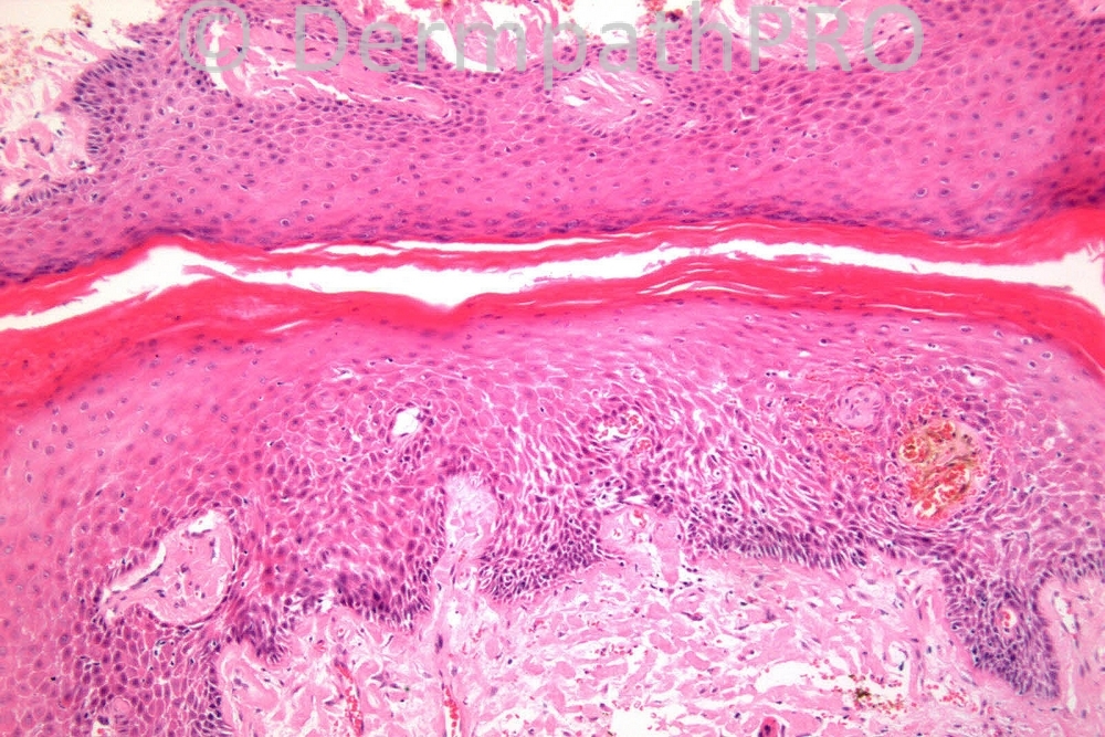
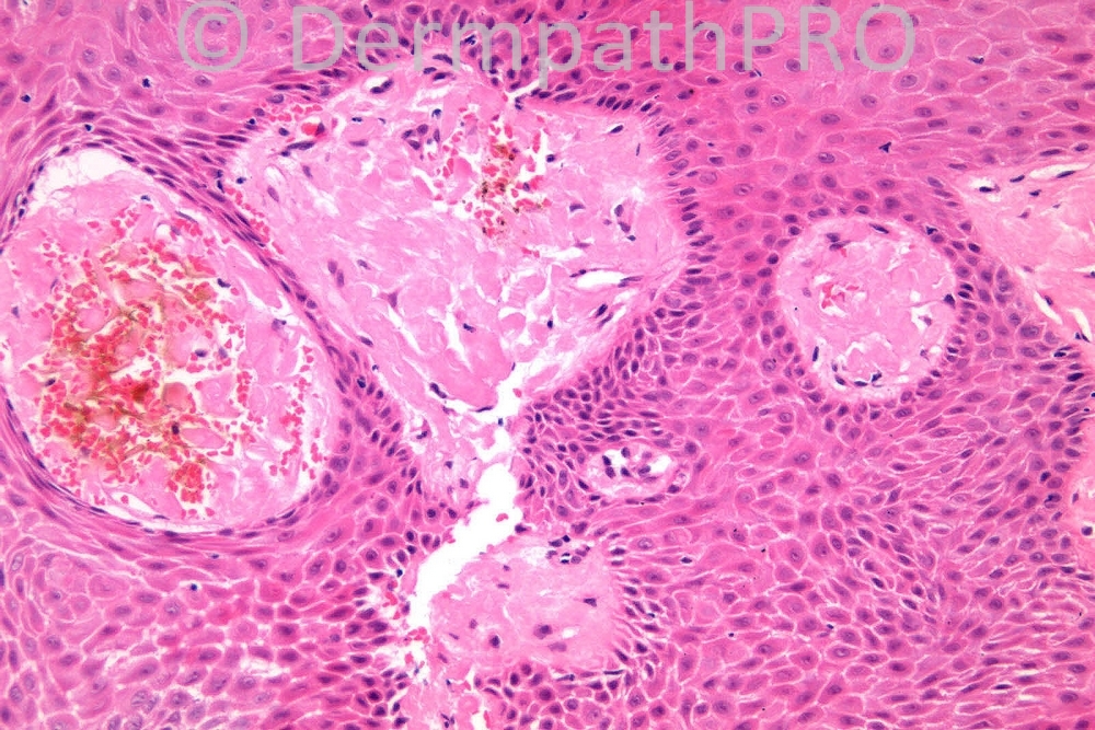
Male 84 years, tight anal stenosis, with sclerotic appearance.


  Report Record

User Feedback


Admin_Dermpath

Posted

[b]Jaime A Mejia, MD - Medicina Diagnóstica (Cali, Colombia) Wrote:[/b]

Erratum: previous post should read, NOT RELATED TO ANY (NOXIOUS) ENTITY ORIGINATED IN THE EPIDERMIS.

[size=2]Submitted on 16/06/2012 04:04[/size] [b]Mahmood - () Wrote:[/b]

Fungal infestation (likely candida)....amyloid deposits, possibly localized, due to chronic irritation.

[size=2]Submitted on 16/06/2012 03:50[/size] [b]Jaime A Mejia, MD - Medicina Diagnóstica (Cali, Colombia) Wrote:[/b]

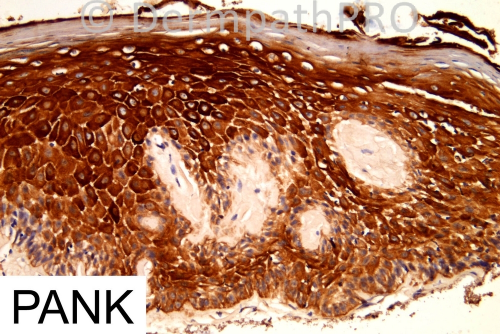
Noh-hereditary organ-limited cutaneous amyloidosis tend to accumulate in the papillary dermis. Systemic amyloidosis tend to affect papillary dermis AND deep dermal structures. CK negativity leads to think of a non-secondary form of amyloidosis related to an entity originated in the epidermis. Schreml, S et al. Eur J Dermatol 2010; 20:152-60

[size=2]Submitted on 15/06/2012 18:33[/size] [b]Richard Carr - Warwick (UK) Wrote:[/b]

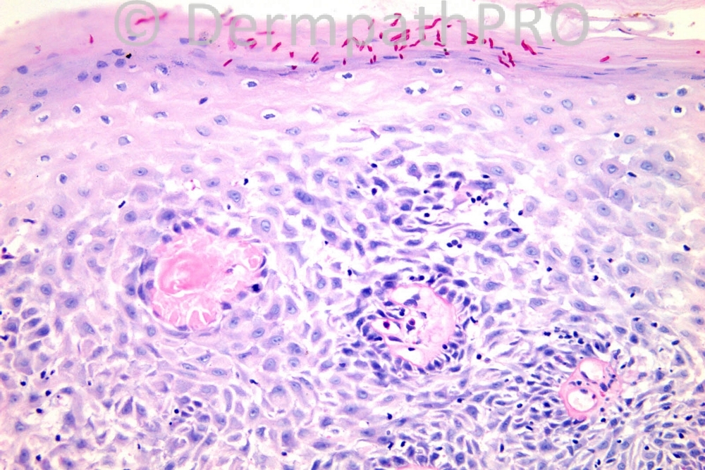
Yes, very well done, candidiasis (mucocutaneous location with pseudohyphae) and amyloidosis. Because the amyloid is pan-keratin negative patient needs investigating for systemic amyloidosis. Cutaneous types of amyloid are usually keratin derived deposits from necrotic keratinocytes (lichen amyloidosis, macular amyloid, cutaneous epithelial tumours notably BCC, SEBK etc).

[size=2]Submitted on 15/06/2012 16:27[/size] [b]Jaime A Mejia, MD - Medicina Diagnóstica (Cali, Colombia) Wrote:[/b]

Primary cutaneous amyloidosis (?), Congo Red stains required. CK-negative deposits, intense CK in epidermis. Was any pruritus ani symptom? Ishii M, Asai Y, Hamada T. Evaluation of cutaneous amyloid employing anti-keratin antibodies and the immunoperoxidase technique (PAP method). Acta Derm Venereol. 1984;64(4):281-5

[size=2]Submitted on 15/06/2012 15:02[/size] [b]Luis Fernando Cardenas Cardona - Centro Dermatologico de Cali (Cali, Colombia) Wrote:[/b]

Systemic Amyloidosis, and fungi

[size=2]Submitted on 15/06/2012 13:57[/size] [b]Azza Mostafa - (Egypt) Wrote:[/b]

Agree systemic Amyloidosis + candidiasis (spores & pseudohyphae)

[size=2]Submitted on 15/06/2012 13:23[/size] [b]Sharmila M - UHCW (UK) Wrote:[/b]

As a dermatology trainee who views these cases with a lot of interest and more often than not, has no clue why others are considering a certain diagnosis in preference to others, I wonder if Dr McKee would consider adding a line or two about the salient features when he gives the answers! This would make this such a great learning opportunity for people like me.

[size=2]Submitted on 15/06/2012 13:08[/size] [b]Rodrigo Restrepo - UPB/CES (Mdlln/COL) Wrote:[/b]

Amyloidosis. Secondary candidiasis

[size=2]Submitted on 15/06/2012 13:05[/size] [b]Yüksel Okumuş - Bursa State Hospital (Turkey) Wrote:[/b]

Candida OK. Subepitelyal amorphous eosinophilic globular material must be amyloid, but needs special stains. All of them ( anal stenosis, candidiasis and secondary amyloidosis )maybe the complications of Crohn disease. PanCK ??

[size=2]Submitted on 15/06/2012 12:37[/size] [b]Mona Abdel Halim - () Wrote:[/b]

Amyloidosis with secondary fungal infection. The presence of hge and the lack of PANK staining favors systemic type.

[size=2]Submitted on 15/06/2012 11:43[/size] [b]Engin Sezer - (Istanbul) Wrote:[/b]

Amyloidosis (possibly systemic -based on lack of CK staining of the amorphous globules-) with superimposing candidal infection.

[size=2]Submitted on 15/06/2012 11:11[/size] [b]Marcela Saeb Lima - INCMNSZ (Mexico City) Wrote:[/b]

Amyloidosis with secondary candidiasis

[size=2]Submitted on 15/06/2012 11:06[/size] [b]I. Abdul-kadir - ST2 - York Hospital (UK) Wrote:[/b]

?Amyloidosis. I'm presuming the fungi are secondary.

[size=2]Submitted on 15/06/2012 10:36[/size]

Share this comment


Link to comment
share_externally


×
×
  • Create New...