Jump to content
In this section we have spot diagnoses posted on a daily basis since June 2010, now over 4000! You can review the archived cases and read the suggested diagnoses by users and the final comment by the contributors.
Case are uploaded each week day by 10 am UK time with the correct diagnosis will generally be posted at 8 pm UK time. Why not view the most recent spot diagnosis and proffer a diagnosis?

Case Number : Case 533 Posted By: Guest

Please read the clinical history and view the images by clicking on them before you proffer your diagnosis.
Submitted Date :
   (0 reviews)

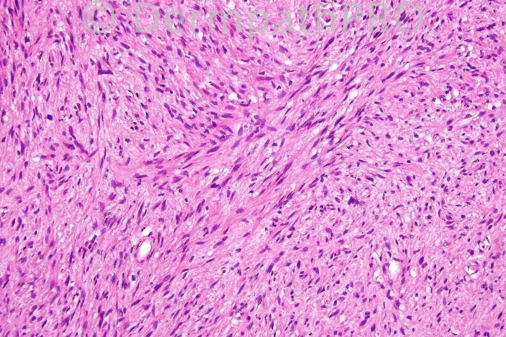
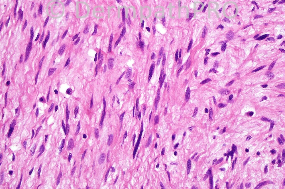
Female 47 years with a firm nodule on her thigh.


  Report Record

User Feedback


Admin_Dermpath

Posted

[b]Jaime A Mejia, MD - Medicina Diagnóstica (Cali, Colombia) Wrote:[/b]

Nice case, very convincing arguments. Thanks Dr. McPhee.

[size=2]Submitted on 25/06/2012 21:33[/size]
[b]Phillip H McKee - Overseasonsultations (Cave Creek, AZ, USA) Wrote:[/b]

Well done all of you who diagnosed MPNST. The mast cells are a clue and there is too much nuclear pleomorphism and hyperchromasia for a neurofibroma in addition to the mitosis. The ill-defined eosinophilic fibrillary cyoplasm is also a diagnostic pointer. The tumor came from a patient with neurofibromatosis type 1.

[size=2]Submitted on 25/06/2012 17:56[/size]
[b]Sasi Attili - Ninewells University Hospital, Dundee (United Kingdom) Wrote:[/b]

I thought leimyosarcoma for this one based on the cigar shaped nuclei. Agree the mast cells are confusing the picture a little bit but the cellular morphology is unlike a NF as i don't see wavy nuclei.

[size=2]Submitted on 25/06/2012 16:10[/size]
[b]Jaime A Mejia, MD - Medicina Diagnóstica (Cali, Colombia) Wrote:[/b]

I have found a report of certain negative correlation between mast cell count and malignancy in soft tissue tumors. In the light of that hypothesis: Could it be a neurofibroma? http://www.ncbi.nlm.nih.gov/pubmed/1594501

[size=2]Submitted on 25/06/2012 15:55[/size]
[b]Jaime A Mejia, MD - Medicina Diagnóstica (Cali, Colombia) Wrote:[/b]

Spindle cutaneous tumor with mast cells, mitotic activity and perinuclear vacuoles. Mast cells can be seen in some ST tumors: synovial sarcoma, neurofibromas, solitary fibrous tumor, GIST, MFH, leiomyosarcomas. My best guess in this case is a cutaneous leiomyosarcoma with mast cells, need to r/o neurofibroma. My panel would include: S-100 prot, HMB-45, SMA, h-caldesmon, desmin, CD34.

[size=2]Submitted on 25/06/2012 15:50[/size]
[b]Zena Slim (SpR Histopath) - Wessex Deanery (QAH Portsmouth) Wrote:[/b]

spindle cell lesion..needs an immuno-panel. Based on morphology my leading differentials are MPNST vs cutaneous Leiomyosarcoma

[size=2]Submitted on 25/06/2012 13:26[/size]
[b]Rodrigo Restrepo - UPB/CES (Mdlln/Col) Wrote:[/b]

Malignant peripheral nerve sheath tumor

[size=2]Submitted on 25/06/2012 12:28[/size]
[b]Marcela Saeb Lima - INCMNSZ (Mexico City) Wrote:[/b]

Agree with Yüksel, malignant peripheral nerve sheath tumor, though there is no pleomorphism there is a mitotic figure.

[size=2]Submitted on 25/06/2012 12:20[/size]
[b]Yüksel Okumuş - Bursa State Hospital (Turkey) Wrote:[/b]

My first impression is malignant peripheral nerve sheath tumor.

[size=2]Submitted on 25/06/2012 12:13[/size]
[b]Engin Sezer - (Istanbul) Wrote:[/b]

Desmoplastic melanoma

[size=2]Submitted on 25/06/2012 11:57[/size]
[b]I. Abdul-kadir - ST2 - York Hospital (UK) Wrote:[/b]

?Dermal leiomyosarcoma.

[size=2]Submitted on 25/06/2012 11:23[/size]

Share this comment


Link to comment
share_externally


×
×
  • Create New...