Jump to content
In this section we have spot diagnoses posted on a daily basis since June 2010, now over 4000! You can review the archived cases and read the suggested diagnoses by users and the final comment by the contributors.
Case are uploaded each week day by 10 am UK time with the correct diagnosis will generally be posted at 8 pm UK time. Why not view the most recent spot diagnosis and proffer a diagnosis?

Case Number : Case 461 Posted By: Guest

Please read the clinical history and view the images by clicking on them before you proffer your diagnosis.
Submitted Date :
   (0 reviews)

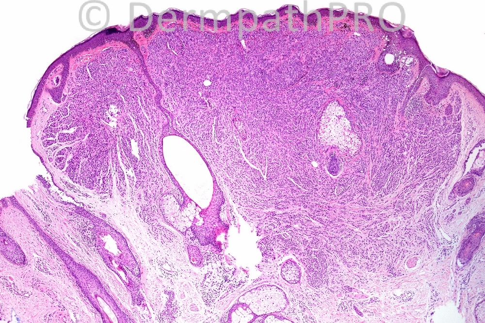
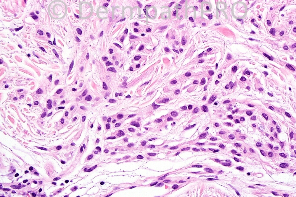
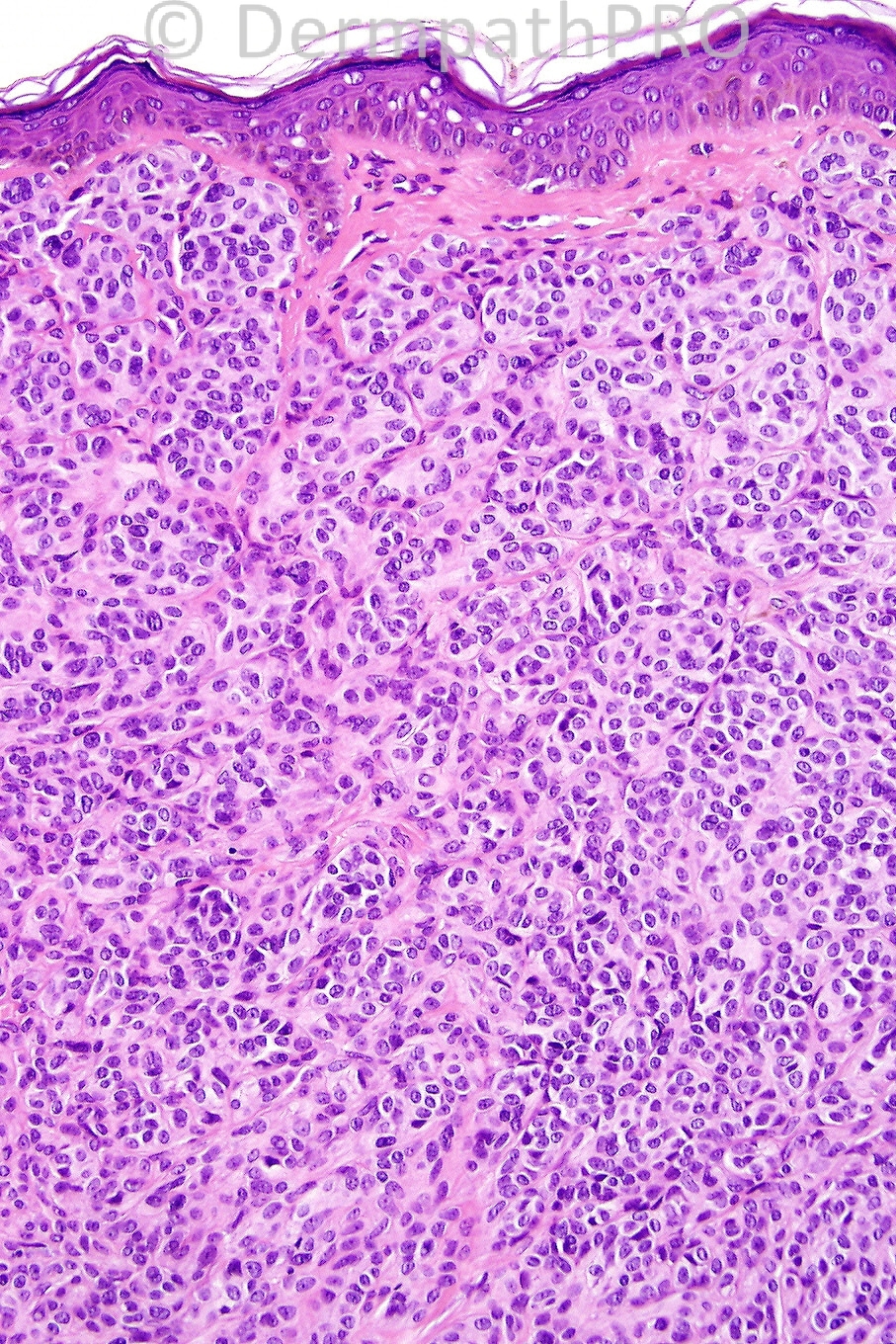
Female 49 years, pigmented lesion on face.


  Report Record

User Feedback


Admin_Dermpath

Posted

[b]Scott Sanders - Warwick Hospital (UK) Wrote:[/b]

The low power appearance is very naevus-like and the deep aspect of the lesion shows maturation with cells streaming off singly and in cords rather than an expansile growth pattern. The presence of a few mitoses is concerning but for me there would have to be significant mitotic activity including atypical mitoses to strongly favour naevoid melanoma over an 'active' naevus.

[size=2]Submitted on 19/03/2012 12:53[/size]
[b]Phillip McKee - Overseas Consultations (Cave Creek, Arizona, USA) Wrote:[/b]

Majority rules okay. Nevoid melanoma is correct. Clues to the diagnosis are subtle lack of maturation, mild pleomorphism, type-A cells at the top and bottom of the lesion, nucleolar prominence and mulitple mitoses (image 5)

[size=2]Submitted on 15/03/2012 19:26[/size]
[b]ANDREW DANCKWERTS - (JOHANNESBURG, RSA) Wrote:[/b]

NEVOID MELANOMA

[size=2]Submitted on 15/03/2012 17:43[/size]
[b]Diana Alvarez - () Wrote:[/b]

Nevoid melanoma.

[size=2]Submitted on 15/03/2012 16:39[/size]
[b]Pablo Ortega - Laboratorio de Patologia Ortega (Loja-Ecuador) Wrote:[/b]

Benign melanocytic nevus

[size=2]Submitted on 15/03/2012 15:30[/size]
[b]Izzat Abdul-kadir - ST2 - York Hospital (UK) Wrote:[/b]

There's symmetry, circumscription and maturation. However, there seems to be "atypia" and dermal mitoses, trumping the latter. Hence, naevoid melanoma.

[size=2]Submitted on 15/03/2012 15:25[/size]
[b]Engin Sezer - Acıbadem University (Istanbul, Turkey) Wrote:[/b]

Congenital melanocytic nevus showing a deep Indian-file pattern, uniform population of type A cells without tendency to form discrete nests superficially and pseudovascular spaces. Mitotic figures remind the motto 'nevi must live'.

[size=2]Submitted on 15/03/2012 14:43[/size]
[b]Rodrigo Restrepo - UPB/CES (Mdlln/Col) Wrote:[/b]

Nevoid Melanoma

[size=2]Submitted on 15/03/2012 14:32[/size]
[b]Yüksel Okumuş - Bursa State Hospital (Turkey) Wrote:[/b]

For differantial dignose nevoid melanoma with the others (mitotically active banal naevi, clonal naevus, congenital naevus etc.) I advise the link bellow. (McKEE's article) http://jcp.bmj.com/content/57/11/1121.full

[size=2]Submitted on 15/03/2012 14:01[/size]
[b]Mona Abdel Halim - () Wrote:[/b]

Nevoid melanoma

[size=2]Submitted on 15/03/2012 13:54[/size]
[b]Sasi Attili - (United Kingdom) Wrote:[/b]

It is certainly a naevoid lesion.This is a tough one for me to make a diagnosis based on 4 images. But, agree that naevoid melanoma is certainly a possibility

[size=2]Submitted on 15/03/2012 13:50[/size]
[b]Marcela Saeb Lima - INCMNSZ (Mexico City) Wrote:[/b]

Nevoid melanoma

[size=2]Submitted on 15/03/2012 13:45[/size]
[b]omar erdogan - () Wrote:[/b]

Mitotically active naevi or congenital nevi

[size=2]Submitted on 15/03/2012 13:42[/size]
[b]Yüksel Okumuş - Bursa State Hospital (Turkey) Wrote:[/b]

Nevoid Melanoma

[size=2]Submitted on 15/03/2012 13:26[/size]
[b]A Bansal - BCU HB (North Wales) Wrote:[/b]

Melanoma, naevoid variant.

[size=2]Submitted on 15/03/2012 13:03[/size]
[b]Wilber J. Martínez C. - CES University (Medellín COL) Wrote:[/b]

Nevoid melanoma

[size=2]Submitted on 15/03/2012 12:53[/size]

Share this comment


Link to comment
share_externally


×
×
  • Create New...