Jump to content
In this section we have spot diagnoses posted on a daily basis since June 2010, now over 4000! You can review the archived cases and read the suggested diagnoses by users and the final comment by the contributors.
Case are uploaded each week day by 10 am UK time with the correct diagnosis will generally be posted at 8 pm UK time. Why not view the most recent spot diagnosis and proffer a diagnosis?

Case Number : Case 499 Posted By: Guest

Please read the clinical history and view the images by clicking on them before you proffer your diagnosis.
Submitted Date :
   (0 reviews)

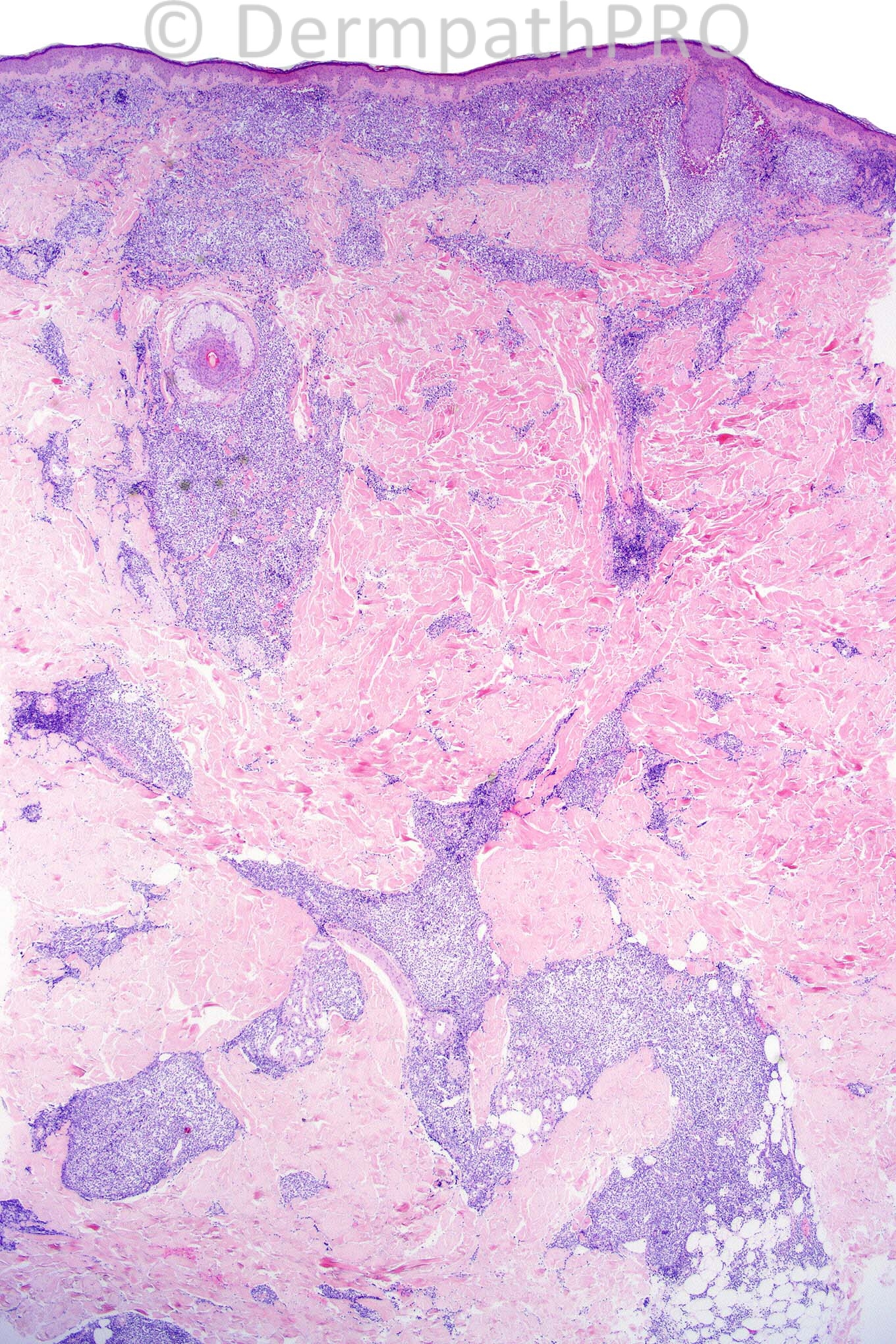
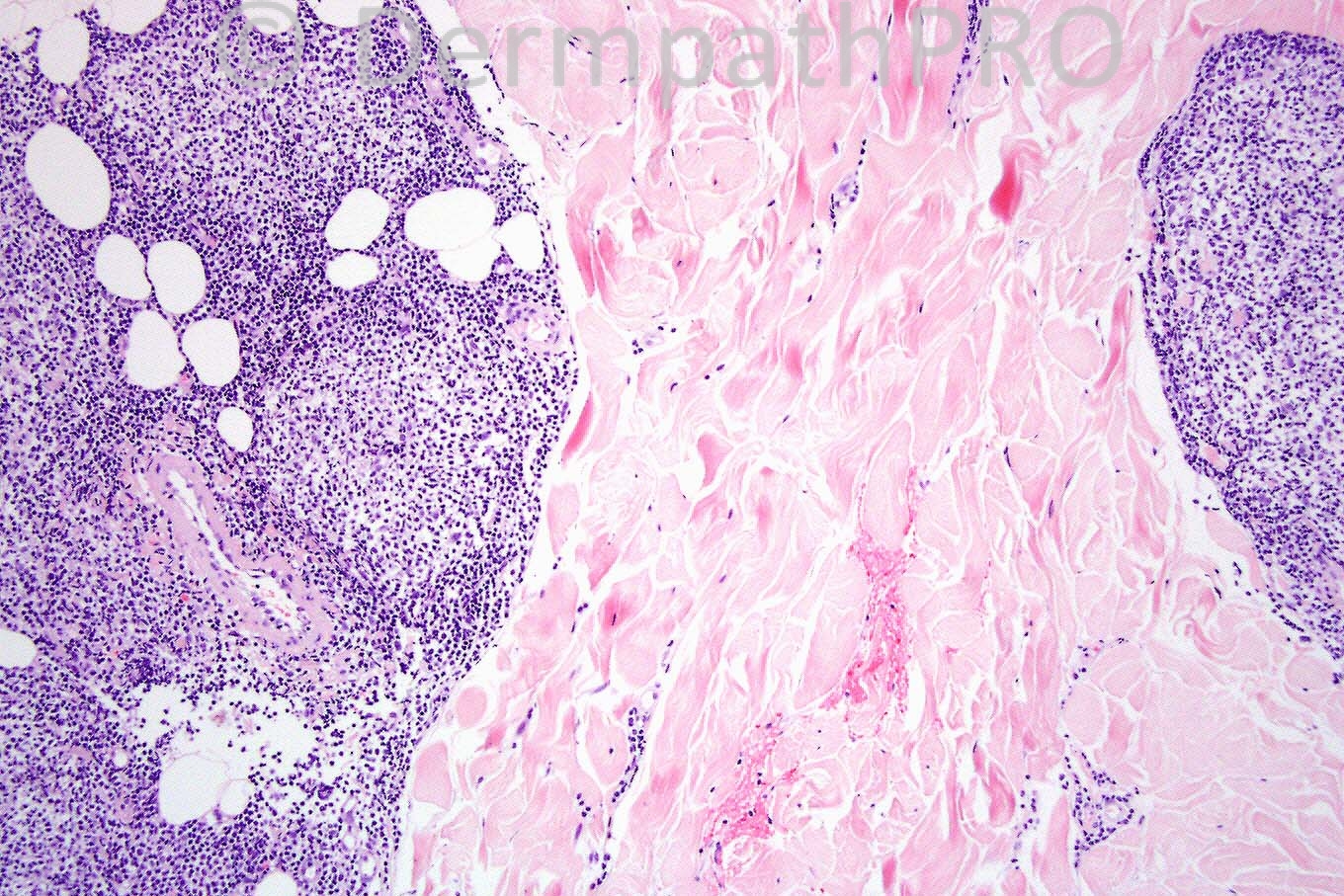
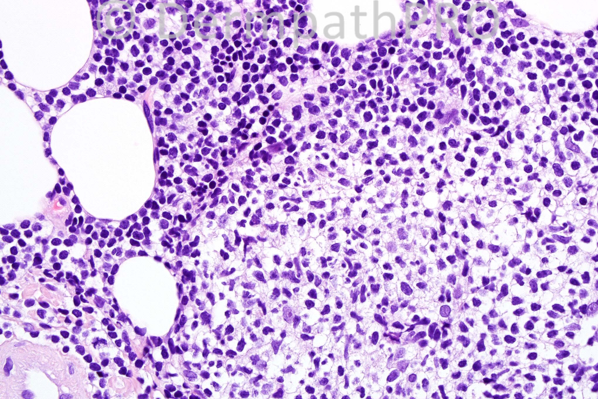
Male 44 years, slightly infiltrated plaque-like skin lesions on the trunk.


  Report Record

User Feedback


Admin_Dermpath

Posted

[b]Richard Carr - Warwick (UK) Wrote:[/b]

Apologies for delay. Well done Rodrigo. This is a case of systemic follicle centre cell lymphoma involving the skin. I am surprised you all found this quite so difficult as the follicles are rather monotonous / relatively inactive looking (quite unlike typical reactive germinal centres) and on these images I cannot see plasma cells that would be expected in the background infiltrate of any reactive process. Also this looks like normal dermal collagen from the back! This would be a good anatomical location for primary cutaneous follicle centre cell lymphoma and clearly immunostains and clinical work-up would be required (I can only presume there was a history of systemic disease).

[size=2]Submitted on 08/05/2012 15:58[/size] [b]Marwa Fawzy - Dermatology Department, Faculty of Medicine, Cairo University (Egypt) Wrote:[/b]

lupus profundus vs lupus profundus like cutaneous T cell lymphoma

[size=2]Submitted on 08/05/2012 15:49[/size] [b]Rodrigo Restrepo - UPB/CES (Mdlln/Col) Wrote:[/b]

B-cutaneous lymphoma

[size=2]Submitted on 08/05/2012 13:44[/size] [b]Mona Abdel Halim - Dermatology Department, Faculty of medicine, Cairo University () Wrote:[/b]

The presence of superficial and deep perivascular and perifollicular infiltrate together with the subcutaneous lobular panniculitis, lymphocyte predominant, and the thick hyalinized vessel in the SC fat as well as the lymphoid follicles in the SC fat with extensive hyalinization of the SC fat and the lack of atypicality in the lymphocytes makes lupus profundus my first diagnosis.

[size=2]Submitted on 08/05/2012 13:42[/size] [b]Marcela Saeb Lima - INCMNSZ (Mexico City) Wrote:[/b]

lupus profundus vs lupus profundus like cutaneous T cell lymphoma

[size=2]Submitted on 08/05/2012 13:34[/size] [b]A Bansal - BCU HB (North Wales) Wrote:[/b]

Diff Dx: lupus erythematosus, lymphoma. Needs immunos.

[size=2]Submitted on 08/05/2012 13:33[/size] [b]Hazem Hamed - Department of Histopathology, Imperial College (LONDON) Wrote:[/b]

I would like to add Tumid LE to the differntial diagnosis of lymphoma and cutaneous lymphoid hyperplasia. Immuno is required.

[size=2]Submitted on 08/05/2012 13:23[/size] [b]Yüksel Okumuş - Bursa State Hospital (Turkey) Wrote:[/b]

B-cutaneous lymphoid hyperplasia. (Borrelia related ?)

[size=2]Submitted on 08/05/2012 13:08[/size] [b]Yüksel Okumuş - Bursa State Hospital (Turkey) Wrote:[/b]

Morphea

[size=2]Submitted on 08/05/2012 12:27[/size] [b]Izzat Abdul-kadir - ST2 - York Hospital (UK) Wrote:[/b]

Lymphoma, for immunos.

[size=2]Submitted on 08/05/2012 12:12[/size]

Share this comment


Link to comment
share_externally


×
×
  • Create New...