Jump to content
In this section we have spot diagnoses posted on a daily basis since June 2010, now over 4000! You can review the archived cases and read the suggested diagnoses by users and the final comment by the contributors.
Case are uploaded each week day by 10 am UK time with the correct diagnosis will generally be posted at 8 pm UK time. Why not view the most recent spot diagnosis and proffer a diagnosis?

Case Number : Case 510 Posted By: Guest

Please read the clinical history and view the images by clicking on them before you proffer your diagnosis.
Submitted Date :
   (0 reviews)

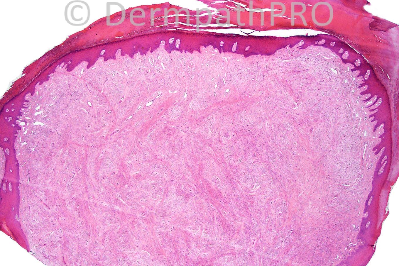
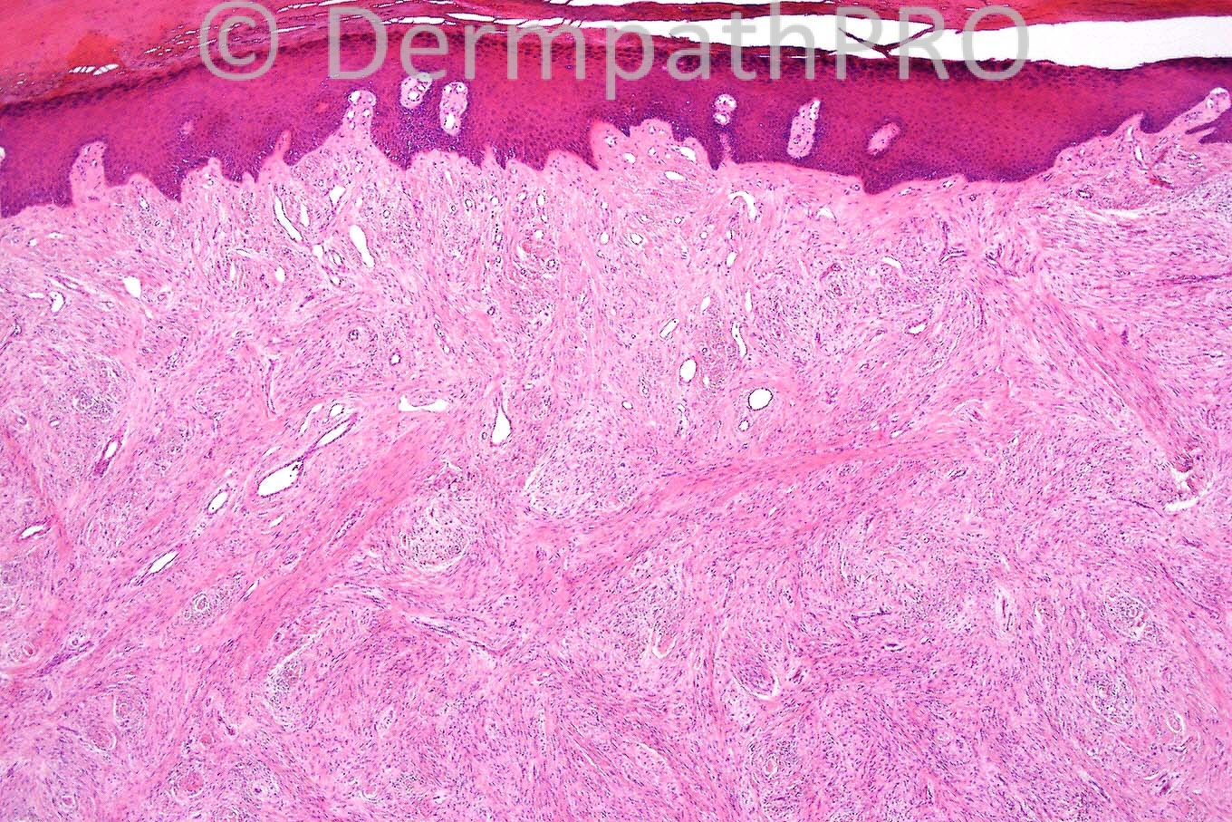
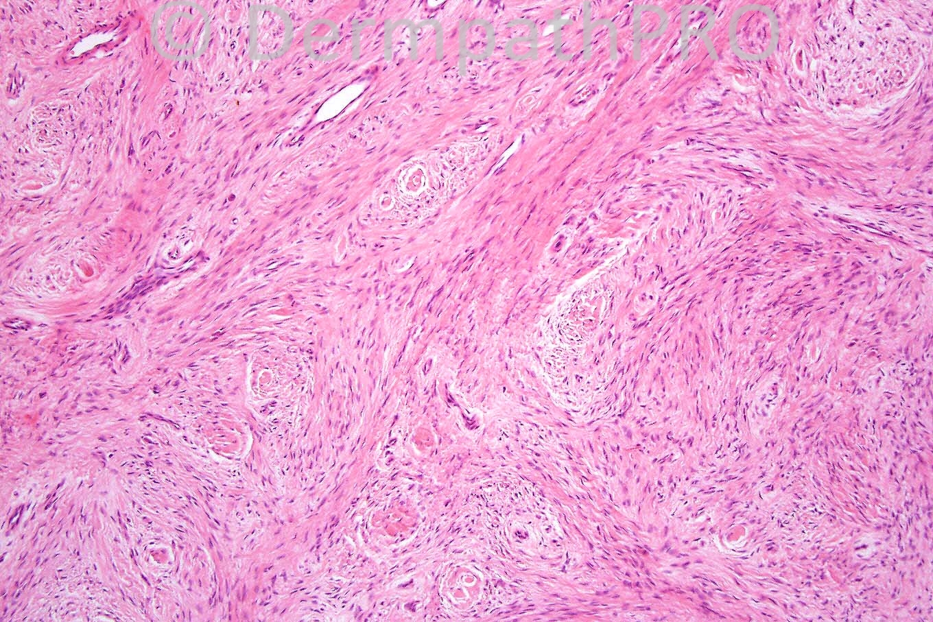
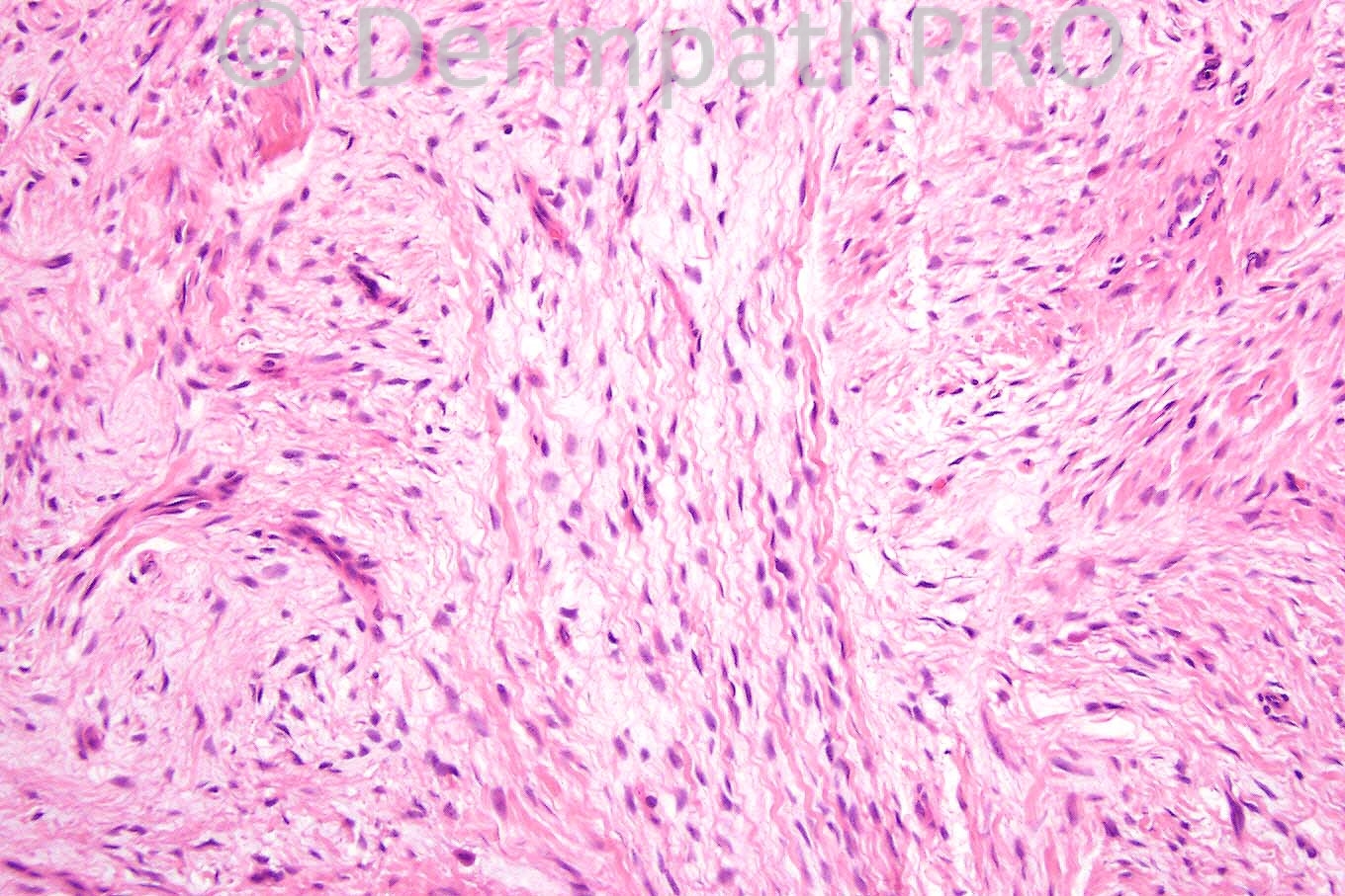
Male 67 years, nodule on left index finger.


  Report Record

User Feedback


Admin_Dermpath

Posted

[b]Phillip H McKee - Overseas Consultations (Cave Creek, ARizona, United States) Wrote:[/b]

Quite a range of diagnoses whichc are mostly very good. The lesion is however a superficial acral fibromyxoma, cellular variant. The alcian blue stain (not shown) dsiclosed an abundance of mucin. Well done Marie amd Eman. Engin, I am sure that your day will come.

[size=2]Submitted on 23/05/2012 21:50[/size]
[b]Azza Mostafa - () Wrote:[/b]

cellular perineuroma, also thought of solitary fibrous tumour

[size=2]Submitted on 23/05/2012 18:14[/size]
[b]Eman El-Nabarawy - () Wrote:[/b]

Superficial acral fibromyxoma.

[size=2]Submitted on 23/05/2012 17:56[/size]
[b]Marie MD - () Wrote:[/b]

I think this has features of cellular fibroma of digits, i.e superficial acral fibromyxoma, cellular variant

[size=2]Submitted on 23/05/2012 17:25[/size]
[b]Mona Abdel Halim - () Wrote:[/b]

Sclerosing perineurioma

[size=2]Submitted on 23/05/2012 16:49[/size]
[b]Engin Sezer - (Istanbul) Wrote:[/b]

Wishing Prof. McKee to say 'Well done Engin'. This would be a great honour for me.

[size=2]Submitted on 23/05/2012 13:29[/size]
[b]Engin Sezer - (Istanbul) Wrote:[/b]

Bland, short, spindle-shaped cells in a storiform growth pattern surrounded by acanthotic and hyperkeratotic epidermis. Sclerosing perineurioma mimicking acquired digital fibrokeratoma. I had the chance to see a slide by Dr. Calonje with such a diagnosis and I have to confess that I first interpreted that as ADF. EMA is heplful for confirmation.

[size=2]Submitted on 23/05/2012 13:24[/size]
[b]Hazem Hamed - Department of Histopathology, Imperial College (LONDON) Wrote:[/b]

Acquired digital fibrokeratoma.

[size=2]Submitted on 23/05/2012 13:08[/size]
[b]Izzat Abdul-kadir - ST2 - York Hospital (UK) Wrote:[/b]

A knuckle pad akin to fibromatosis.

[size=2]Submitted on 23/05/2012 12:55[/size]

Share this comment


Link to comment
share_externally


×
×
  • Create New...