Jump to content
In this section we have spot diagnoses posted on a daily basis since June 2010, now over 4000! You can review the archived cases and read the suggested diagnoses by users and the final comment by the contributors.
Case are uploaded each week day by 10 am UK time with the correct diagnosis will generally be posted at 8 pm UK time. Why not view the most recent spot diagnosis and proffer a diagnosis?

Case Number : Case 631 - 8 Nov Posted By: Guest

Please read the clinical history and view the images by clicking on them before you proffer your diagnosis.
Submitted Date :
   (0 reviews)

Male 60 years lesion on upper abdomen.


  Report Record

User Feedback


Iskander H. Chaudhry

Posted

Please take a moment to check the [url="https://dermpathpro.com/difficult_opinion_cases_1/_/case-do011-r13?&do=comments#commentsStart"]new difficult opinion case[/url] and post your views on it.

Share this comment


Link to comment
share_externally

Guest Dr Engin Sezer

Posted

CN with mild/moderate cytological and architectural atypia

Share this comment


Link to comment
share_externally

Favour a dysplastic naevus

Share this comment


Link to comment
share_externally

Guest Gulerkin

Posted

Compound dyslastic nevus with mild/moderate atypia

Share this comment


Link to comment
share_externally

Sasi Attili

Posted

I would go with Marcela on this one.

Share this comment


Link to comment
share_externally

Guest Rodrigo Restrepo

Posted

Recurrent nevus

Share this comment


Link to comment
share_externally

Guest Robledo F. Rocha

Posted

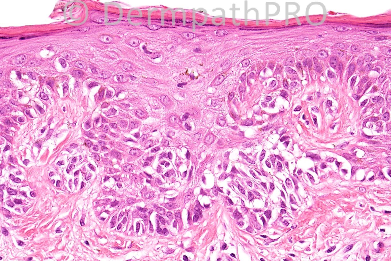
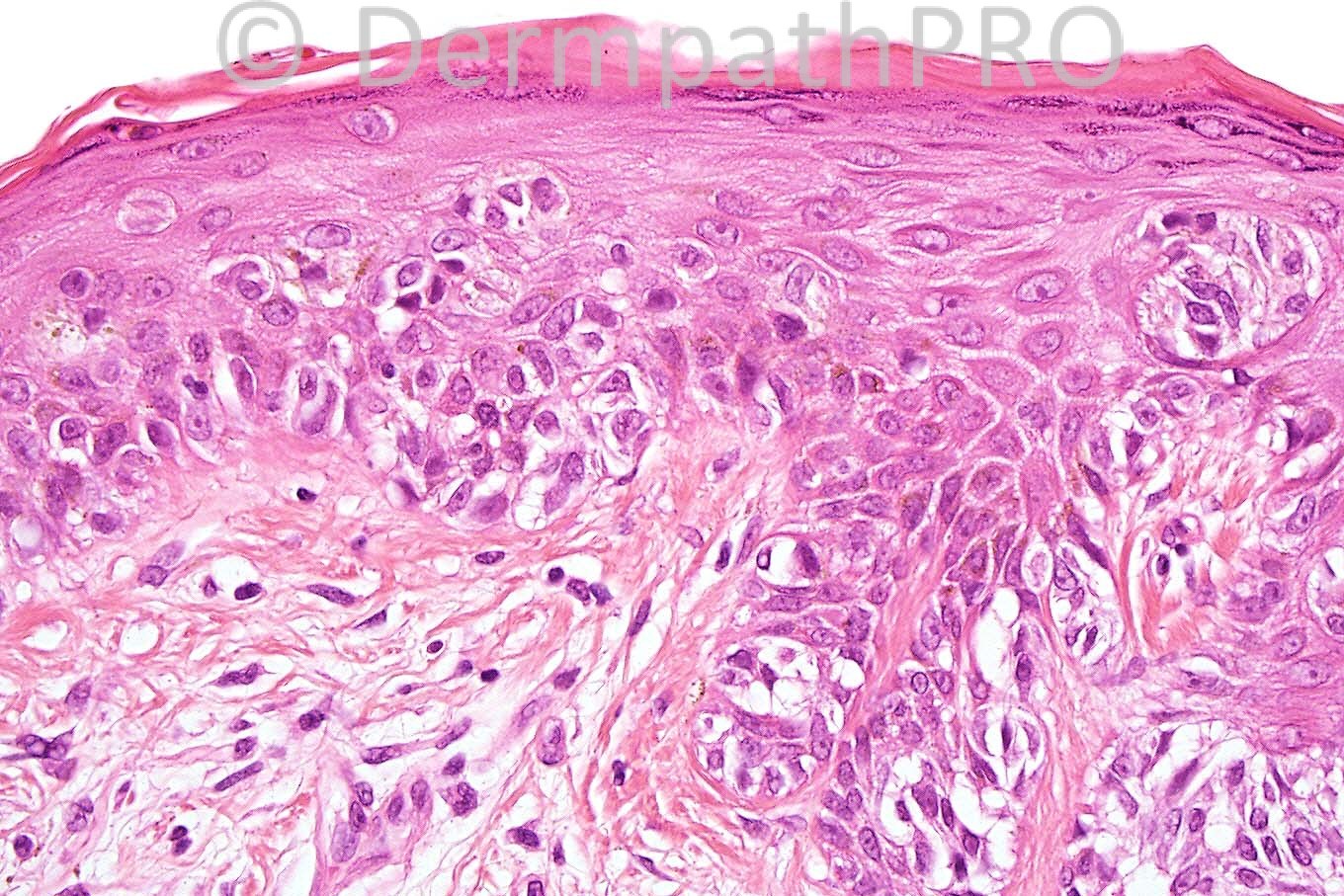
I think it’s a dysplastic nevus. There's architectural disorder (junctional shoulder extending laterally beyond the papillary dermal component; lentiginous melanocytic proliferation and irregular junctional nesting, the last one characterized by variation in size, shape, and location on the sides and between rete ridges with bridging). There's also random cytologic atypia, and host response in the form of lamellar fibroplasia.
The pagetoid spread is so miminal to be worthy of calling the lesion a melanoma in situ.

Share this comment


Link to comment
share_externally

Dr. Mona Abdel-Halim

Posted

Severly dysplastic nevus.

Share this comment


Link to comment
share_externally

Dysplastic naevus. I would not go beyond moderate.

Share this comment


Link to comment
share_externally

Dr. Phillip McKee

Posted

Melanocytic lesions continue to be a problem for you all. The first (low power) view shows that the lesion has a lentiginous growth pattern. The third image shows pagetoid spread (which is not allowed in a dysplastic nevus) and in the 4th image, the dermal nests are larger than the junctional ones (a clear indicator of melanoma). The diagnosis is melanoma and it has been subtyped as the lentiginous variant which is quite different from lentigo maligna (melanoma) and is commonly misdiagnosed as a dysplastic nevus as there is certainly some histological overlap. There is not an awful lot of literaure on the entity.

Share this comment


Link to comment
share_externally

I did mentioned melanoma, not the lentiginous variant, though.

Share this comment


Link to comment
share_externally


×
×
  • Create New...