Jump to content
In this section we have spot diagnoses posted on a daily basis since June 2010, now over 4000! You can review the archived cases and read the suggested diagnoses by users and the final comment by the contributors.
Case are uploaded each week day by 10 am UK time with the correct diagnosis will generally be posted at 8 pm UK time. Why not view the most recent spot diagnosis and proffer a diagnosis?

Case Number : Case 640 - 21 Nov Posted By: Guest

Please read the clinical history and view the images by clicking on them before you proffer your diagnosis.
Submitted Date :
   (0 reviews)

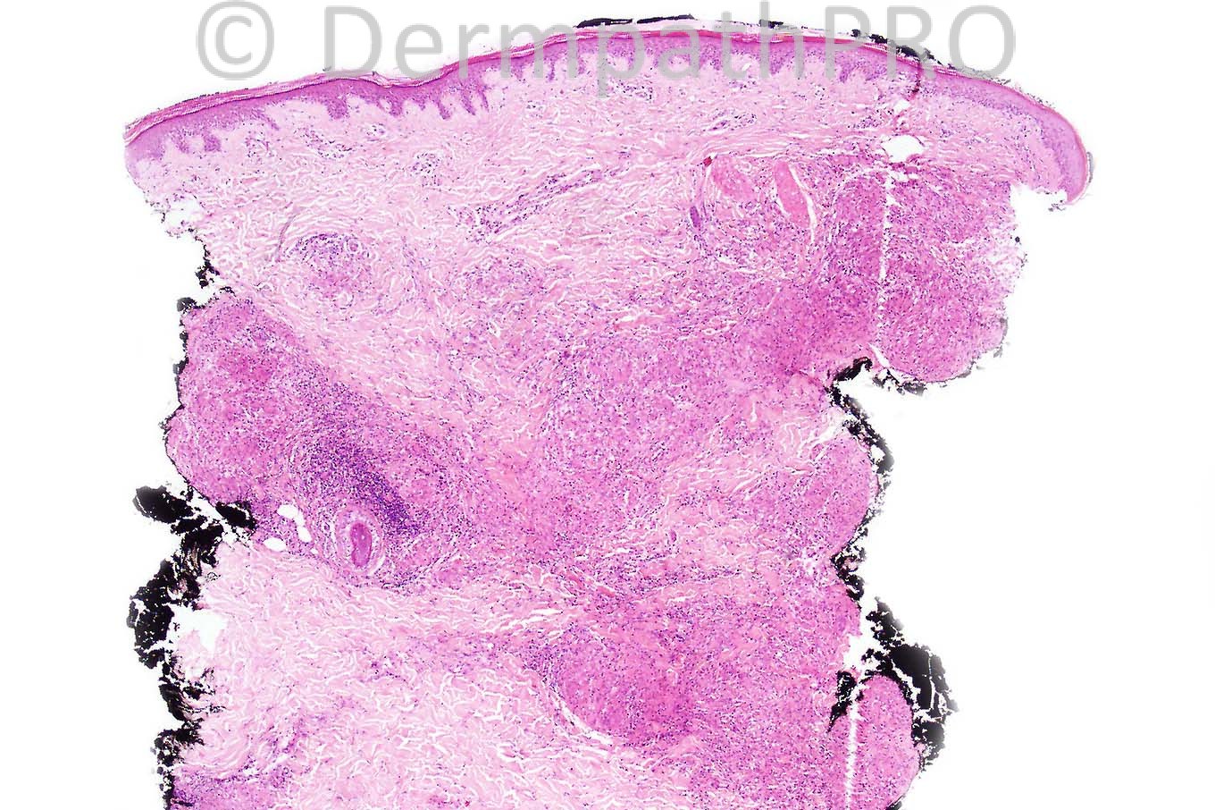
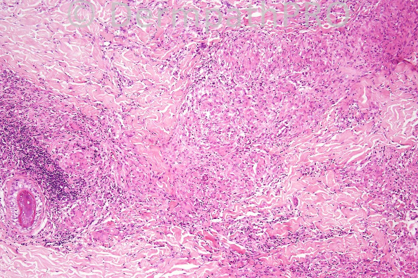
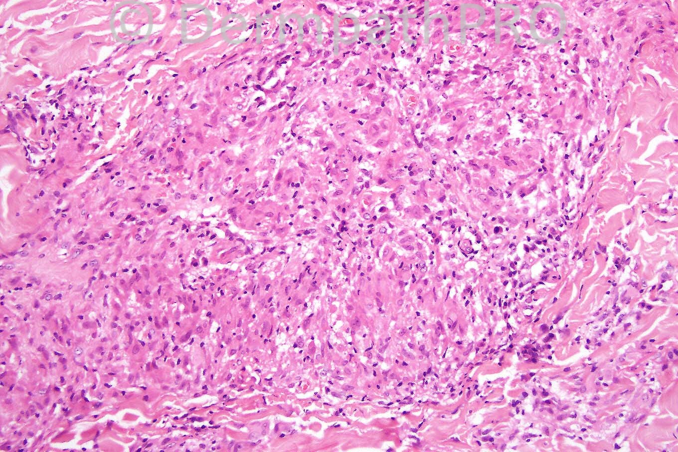
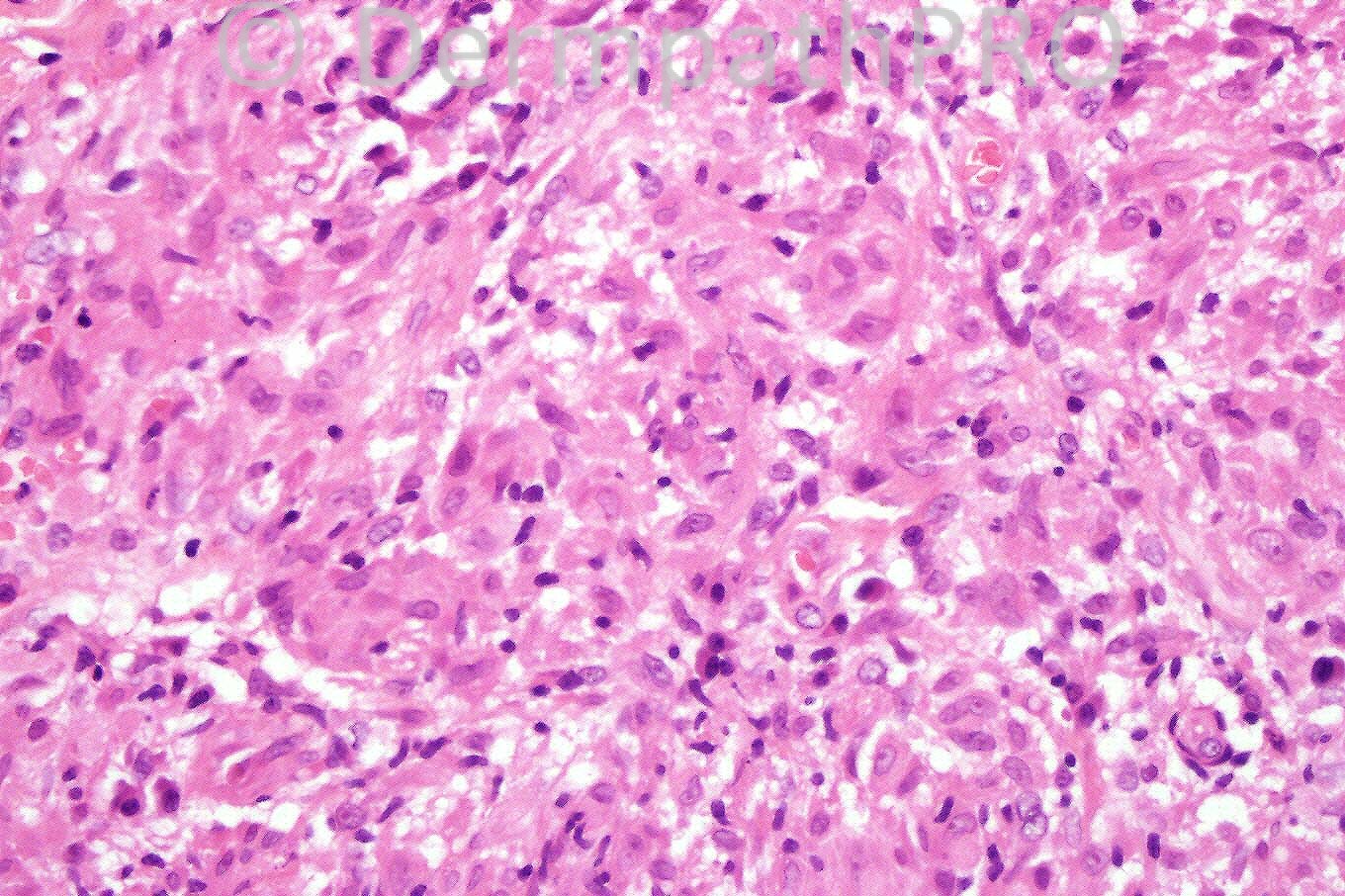
Female 42 years with erythematous plaques on the face and trunk.


  Report Record

User Feedback


Guest Hazem Hamed

Posted

Non necrotizing sarcoid-like granulomata. Special stains (giemsa, Grocott, WS, ZN, DPAS)to exclude infectious aetiology, polarized light to exlude FB. Then correlation with clinical history and radiology (chest) for Sarcoidosis. Is there a history of Crohn's disease?. Cutaneous Crohn's sometimes preceeds GI involvement.

Share this comment


Link to comment
share_externally

Guest Dr Engin Sezer

Posted

My first impression is leprosy based on bubbly ctoplasm of the granulomatous infiltrate, hence Fite stain first..

Share this comment


Link to comment
share_externally

Periadnexal granuloma, foamy pale hitios,++plasma cells ??leprosy

Share this comment


Link to comment
share_externally

Guest Rodrigo Restrepo

Posted

Agree, Hansen's disease, maybe borderline tuberculoid.

Share this comment


Link to comment
share_externally

Guest Robledo F. Rocha

Posted

It seems to be borderline leprosy, an infectious disease that is unfortunately endemic here in Brazil. There are collections of epithelioid macrophages without the formation of well-defined granulomas. Some of these cells have foamy cytoplasm plenty of fragmented acid-fast bacilli, and they are called Virchow cells There are also scanty lymphocytes, but no Langhans giant cells. The granulomas are stretched out because they follow along the neurovascular bundles.

Share this comment


Link to comment
share_externally

Dr. Mona Abdel-Halim

Posted

I will go also for leprosy. Borderline tuberculoid/ Borderline leprosy.

Share this comment


Link to comment
share_externally

Eman El-Nabarawy

Posted

I can see sarcoidal granulomas formed of epithellioid cells with giant cells (fig 1) and asteroid body (fig 4). So I will favor sarcoidosis. Borderline tuberculoid leprosy will show tuberculoid granulomas rich in lymphocytes and borderline lepromatous leprosy will not show epithelioid cells or giant cells. There is a variant of sarcoidosis rich in plasma cells.

Share this comment


Link to comment
share_externally

Arif Usmani

Posted

I agree with predominant opinion. However late secondary syphilis may also have show granulomata and lymphoplasmacytic infiltrate but I do not see significant epidermal changes.

Share this comment


Link to comment
share_externally

Dr. Phillip McKee

Posted

All of your differential diagnoses are excellent. The final diagnosis was sarcoidosis. Other than the perifollicular chronic inflammation, the granulomata are naked- a good clue for sarcoid. Never the less special stains must always be done to exclude an infectious etiology.

Share this comment


Link to comment
share_externally

Arif Usmani

Posted

Excellent point, if one removes the perifollicular inflammatory focus from the micrograph then the first thing that comes to mind would be sarcoid. This case is a good exercise to learn to delete the distractor!

Share this comment


Link to comment
share_externally


×
×
  • Create New...