Jump to content
In this section we have spot diagnoses posted on a daily basis since June 2010, now over 4000! You can review the archived cases and read the suggested diagnoses by users and the final comment by the contributors.
Case are uploaded each week day by 10 am UK time with the correct diagnosis will generally be posted at 8 pm UK time. Why not view the most recent spot diagnosis and proffer a diagnosis?

Case Number : Case 641 - 22 Nov Posted By: Guest

Please read the clinical history and view the images by clicking on them before you proffer your diagnosis.
Submitted Date :
   (0 reviews)

Female 44 years with scaly plaques on the knees.


  Report Record

User Feedback


prurigo nodularis(atopic- many eos.)

Share this comment


Link to comment
share_externally

Guest Hazem Hamed

Posted

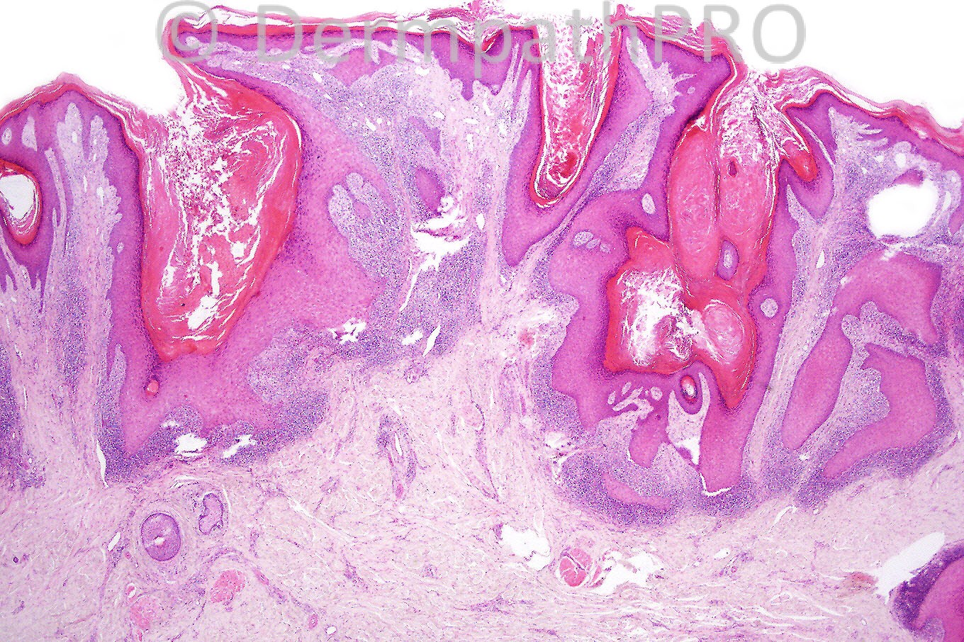
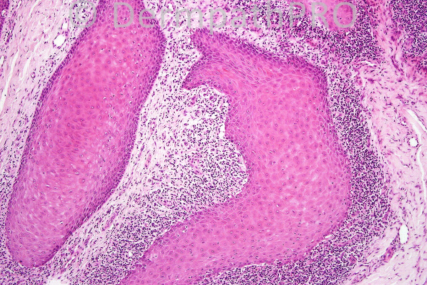
Hypertrophic lichen planus. Hypergranulosis, acanthosis, squamatization of basal epidermis, band-like infiltrate mostly arranged at the tips of the rete ridges.

Share this comment


Link to comment
share_externally

Guest Dr Engin Sezer

Posted

Prurigo/picker's nodule based on hairy-palm sign, hypergranulosis, slight spongiosis and lack of interphase changes. Conspicious eosinophils may occasionaly be observed in prurigo.

Share this comment


Link to comment
share_externally

Guest Dr Engin Sezer

Posted

However it is difficult to rule out pemphigoid nodularis without DIF. Recently I had a patient with that diagnosis and read the article from St. John's -which I miss so much- with great interest.

Share this comment


Link to comment
share_externally

Guest Robledo F. Rocha

Posted

I think it’s a hypertrophic lichen planus, typically located in the lower limb.
There are changes (psoriasiform hyperplasia of the epidermis and thickened papillary dermis) superposed to a classic lesion of lichen planus due to persistent and vigorous rubbing.
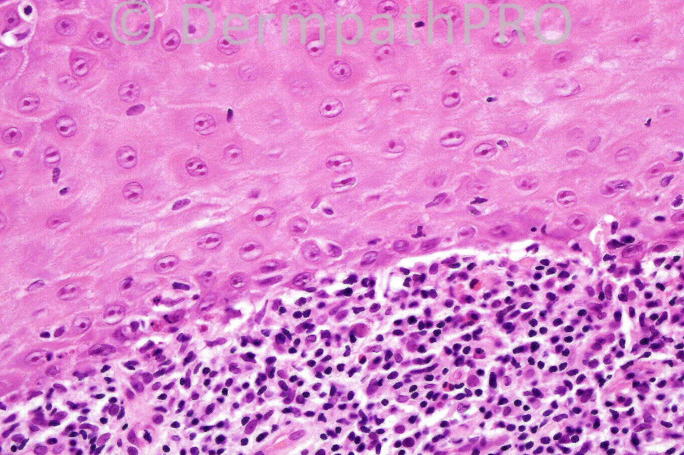
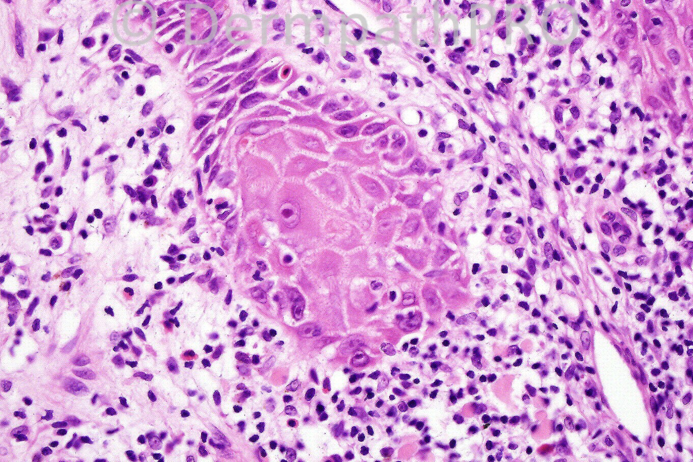
Features of lichen planus that I found are wedge shape hypergranulosis related to the acrosyringia and acrotrichia (Fig. 1), and epidermal basal cell damage more prominent in the tips of the rete ridges (Fig. 4). There’s also a band-like infiltrate of lymphocytes, and a few eosinophils and plasma cells, the last two types of inflammatory cells suggesting ingestion of beta-blockers.

Share this comment


Link to comment
share_externally

Dr. Phillip McKee

Posted

Hypertrophic lichen planus it is. Against picker's nodule are the band-like infiltrate, basal cell liquifactive degeneration and cytoid bodies seen in Fig 4.

Share this comment


Link to comment
share_externally

Guest Raquel Fernandes

Posted

Dr . Phillip, did you rule out lichenoid keratosis (maybe because of the clinic presentation )?

Share this comment


Link to comment
share_externally


×
×
  • Create New...