Jump to content
In this section we have spot diagnoses posted on a daily basis since June 2010, now over 4000! You can review the archived cases and read the suggested diagnoses by users and the final comment by the contributors.
Case are uploaded each week day by 10 am UK time with the correct diagnosis will generally be posted at 8 pm UK time. Why not view the most recent spot diagnosis and proffer a diagnosis?

Case Number : Case 642 - 23 Nov Posted By: Guest

Please read the clinical history and view the images by clicking on them before you proffer your diagnosis.
Submitted Date :
   (0 reviews)

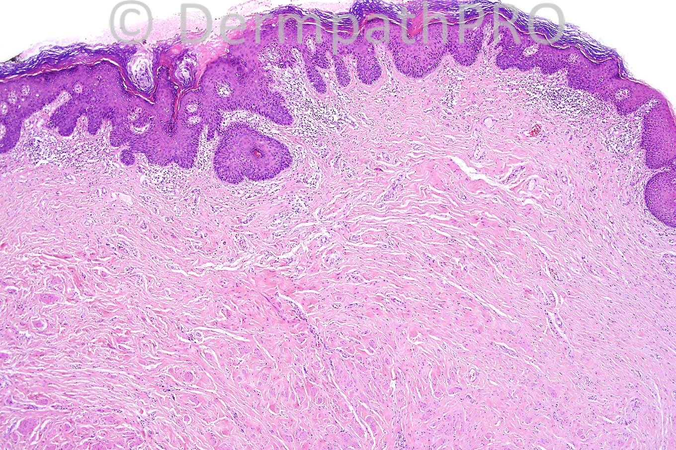
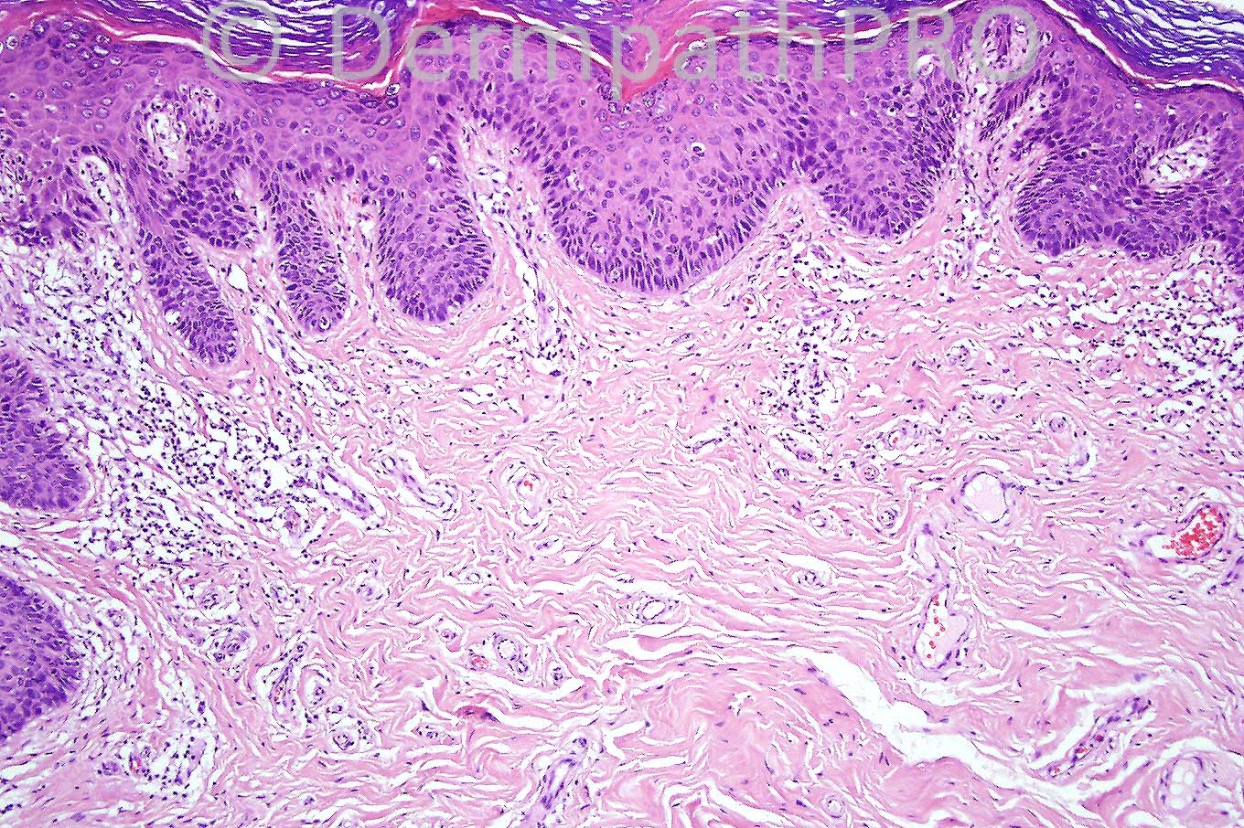
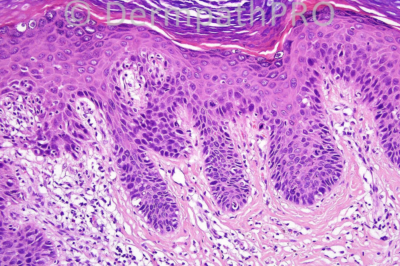
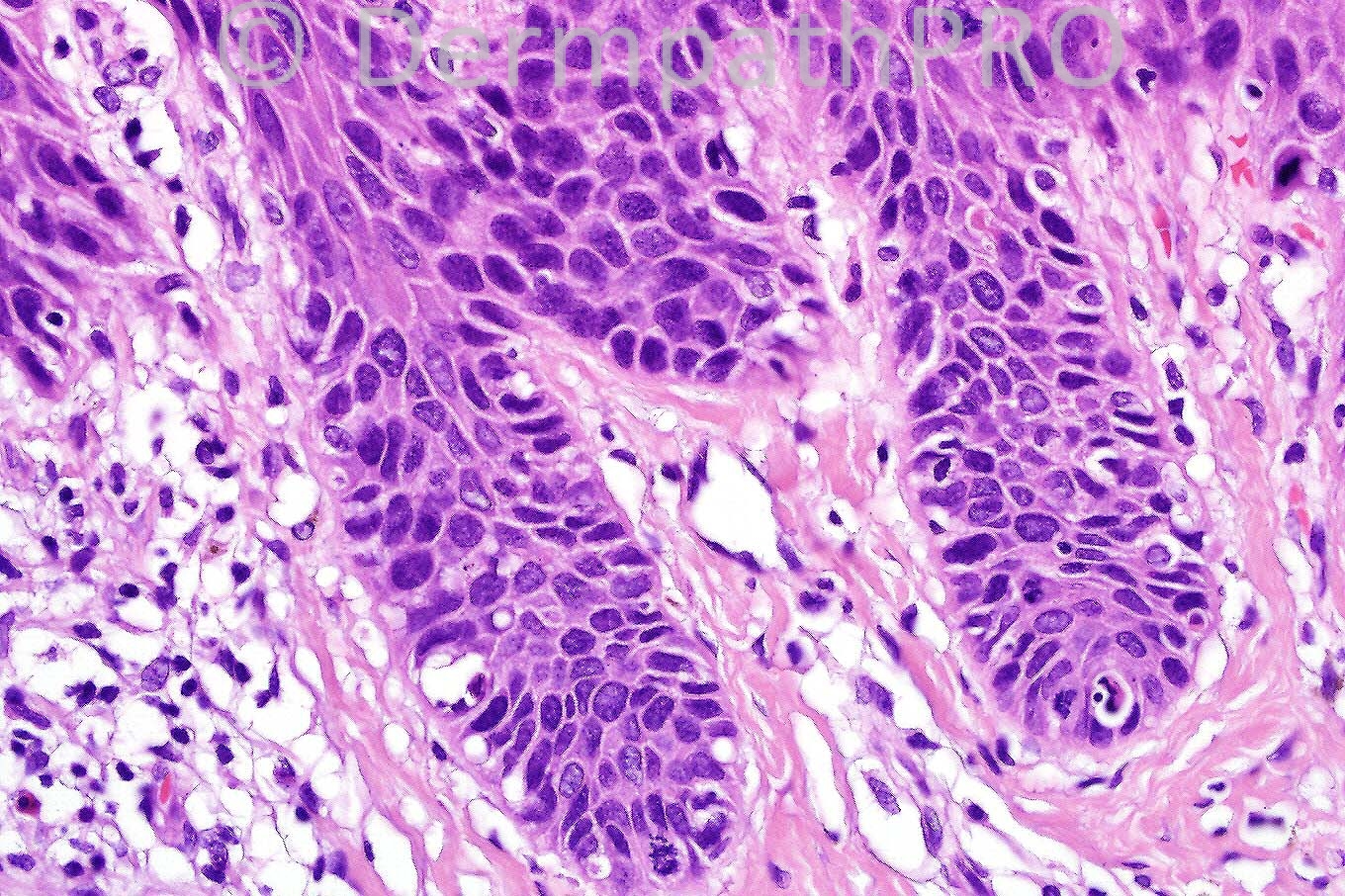
Female 62 years.  History of plaque on vulva.


  Report Record

User Feedback


Guest Hazem Hamed

Posted

VIN (grade 1-2) with features suggestive of HPV related changes. There is no invasive malignancy.

Share this comment


Link to comment
share_externally

Sasi Attili

Posted

This looks like differentiated VIN (greater risk of invasion, though none obvious here), but agree that there seems to be koilocytic change suggestive of HPV infection.

Share this comment


Link to comment
share_externally

Guest Dr Engin Sezer

Posted

Agree with VIN with HPV aetiology

Share this comment


Link to comment
share_externally

Guest Rodrigo Restrepo

Posted

[color=#1C2837][font=arial, verdana, tahoma, sans-serif][size=4]Differentiated VIN[/size][/font][/color]

Share this comment


Link to comment
share_externally

Dr. Mona Abdel-Halim

Posted

HPV associated VIN

Share this comment


Link to comment
share_externally

Dr. Phillip McKee

Posted

I am posting the diagnosis early today as I have to go out shortly. I agree with the diagnosis of HPV associated VIN (II). This is not differentiated VIN which is characterized by cytological atypia affecting the basal layer associated with dyskeratosis. The epidermis differentiates normally which is clearly not the case in this lesion. Many thanks for your comments this week. Have a great weekend.

Share this comment


Link to comment
share_externally


×
×
  • Create New...