Jump to content
In this section we have spot diagnoses posted on a daily basis since June 2010, now over 4000! You can review the archived cases and read the suggested diagnoses by users and the final comment by the contributors.
Case are uploaded each week day by 10 am UK time with the correct diagnosis will generally be posted at 8 pm UK time. Why not view the most recent spot diagnosis and proffer a diagnosis?

Case Number : Case 644 - 27 Nov Posted By: Guest

Please read the clinical history and view the images by clicking on them before you proffer your diagnosis.
Submitted Date :
   (0 reviews)

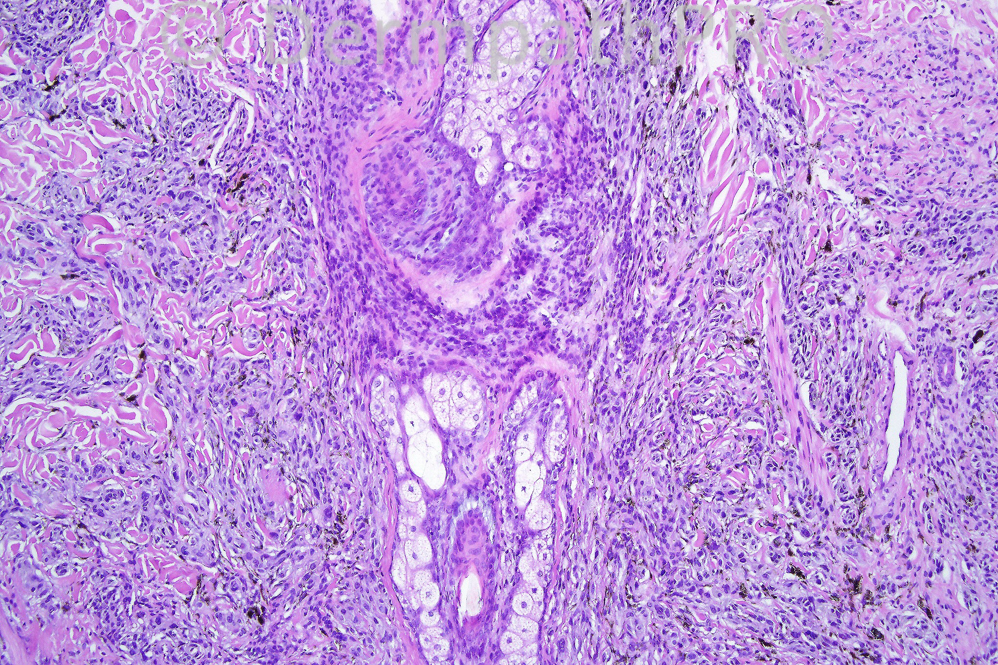
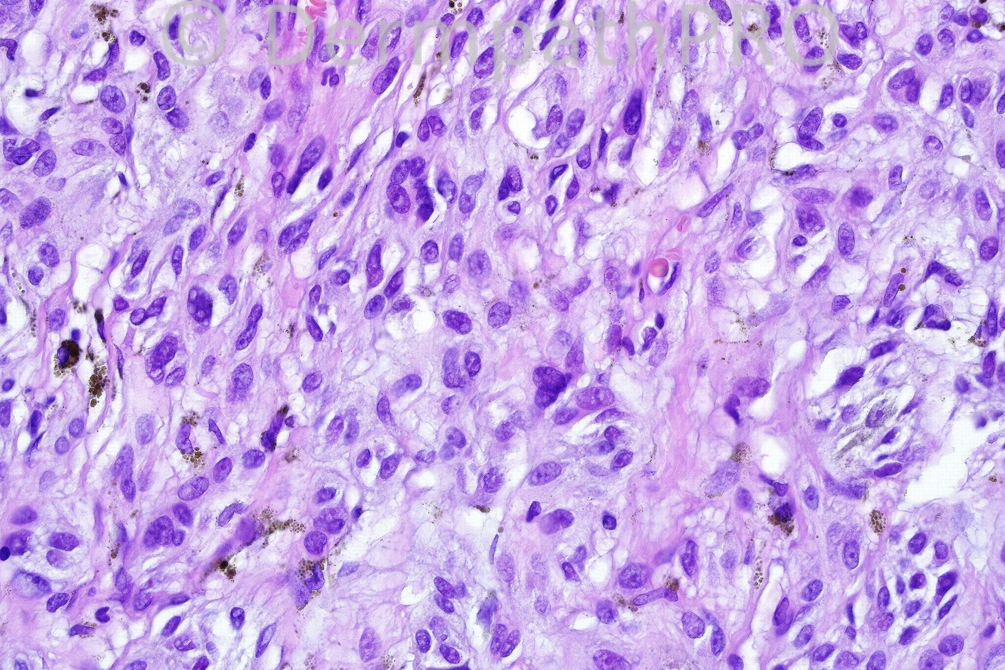
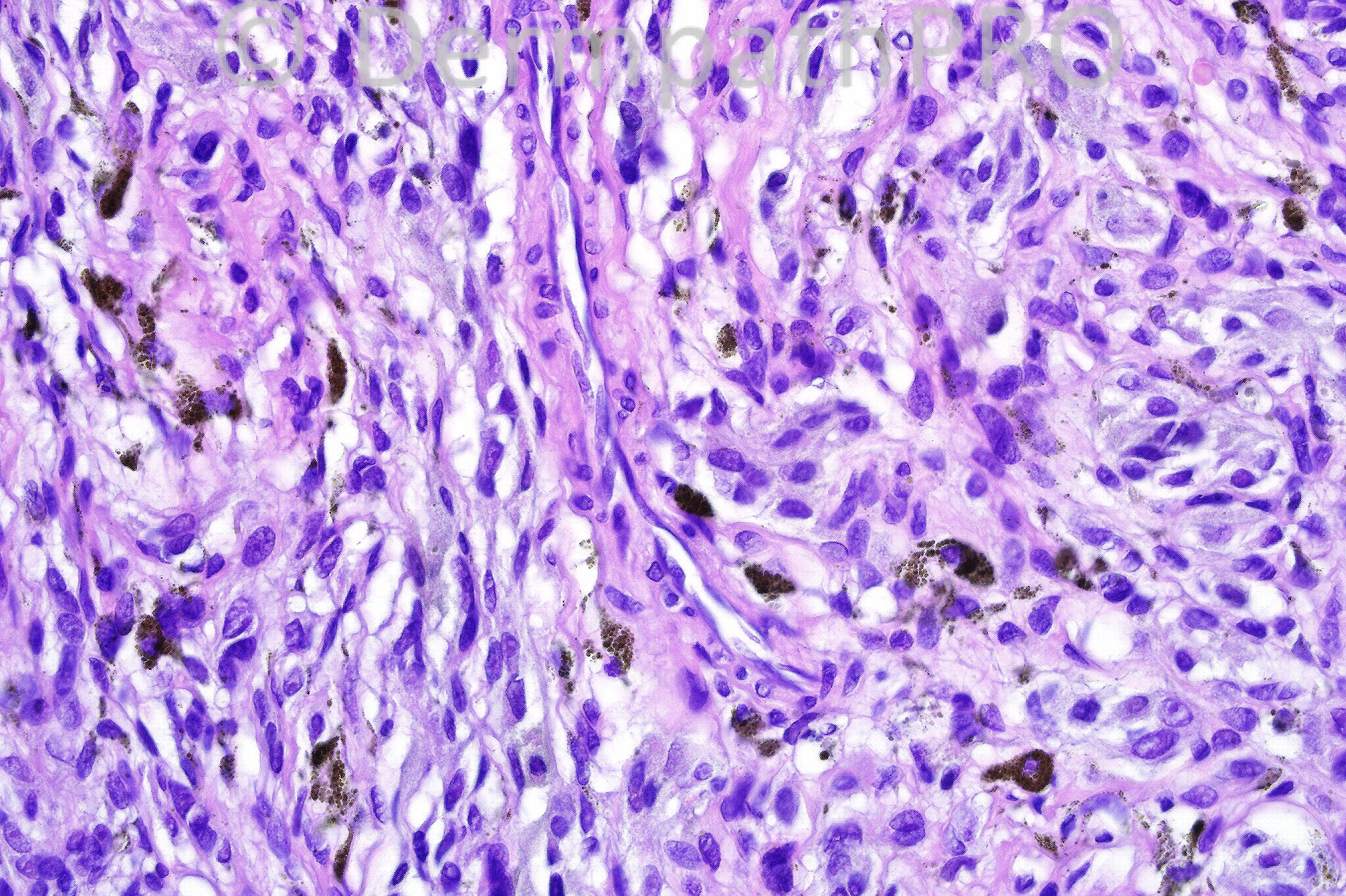
Female 42 years with a irregularly pigmented lesion on the arm. 


  Report Record

User Feedback


Guest Hazem Hamed

Posted

I am not sure if the first image shows dermal nest of nevus cells. Higher power of this areas in not shown. There is no junctional component as far as I can see. The dermis is not sclerotic. Cellular blue nevus versus Deep penetrating nevus.

Share this comment


Link to comment
share_externally

BLUE NEVUS area of uninvolved dermis, spindle shaped cells, excess melanin,ill defined

Share this comment


Link to comment
share_externally

Guest Dr Engin Sezer

Posted

The cells appeared to me too pleomorphic and hyperchromatic for a banal cellular blue neavus, however I couldn't catch a mitotic figure though, hence I'm not comfortable regarding the possibility of a cellular blue naevus-like melanoma (i.e. melanoma arising on a congenital naevus).

Share this comment


Link to comment
share_externally

Arif Usmani

Posted

Architecturally the lesion appears to be a deep penetrating nevus or a combined nevus however due to cytologic atypia, irregular melanization and some degree of pleomorphism I would examine the lesion closely for mitotic activity to rule out a possibility of melanoma arising within a nevus. There is also asymmetry as the upper right side of the lesion does not show more differentiated type “B” melanocytes.

Share this comment


Link to comment
share_externally

Dr. Phillip McKee

Posted

This is a combined banal and deep penetrating nevus. The low power view shows the wedge-shaped growth pattern with banal nevus at the top. Top right emphasizes the folliculocentricity. Bottom left is from near the top of the lesion showing a more epithelioid cell population whereas the bottom right shows the spindled cell population in the deep part of the lesion with some dendritic cells. I classify deep penetrating nevus as a variant of blue nevus so the other diagnoses of blue nevus were not so far off the mark.

Share this comment


Link to comment
share_externally

Guest Robledo F. Rocha

Posted

My hypothesis is combined nevus. This wedge-shaped lesion consists of a fascicular proliferation of pigmented spindle cells with an overlying intradermal melanocytic nevus.

Share this comment


Link to comment
share_externally

Arif Usmani

Posted

Please correct the title to read Case 644 (it is labeled as 444).

Share this comment


Link to comment
share_externally

Guest MarieMD

Posted

combined nevus, congenital and blue

Share this comment


Link to comment
share_externally


×
×
  • Create New...