Jump to content
In this section we have spot diagnoses posted on a daily basis since June 2010, now over 4000! You can review the archived cases and read the suggested diagnoses by users and the final comment by the contributors.
Case are uploaded each week day by 10 am UK time with the correct diagnosis will generally be posted at 8 pm UK time. Why not view the most recent spot diagnosis and proffer a diagnosis?

Case Number : Case 645 - 28 Nov Posted By: Guest

Please read the clinical history and view the images by clicking on them before you proffer your diagnosis.
Submitted Date :
   (0 reviews)

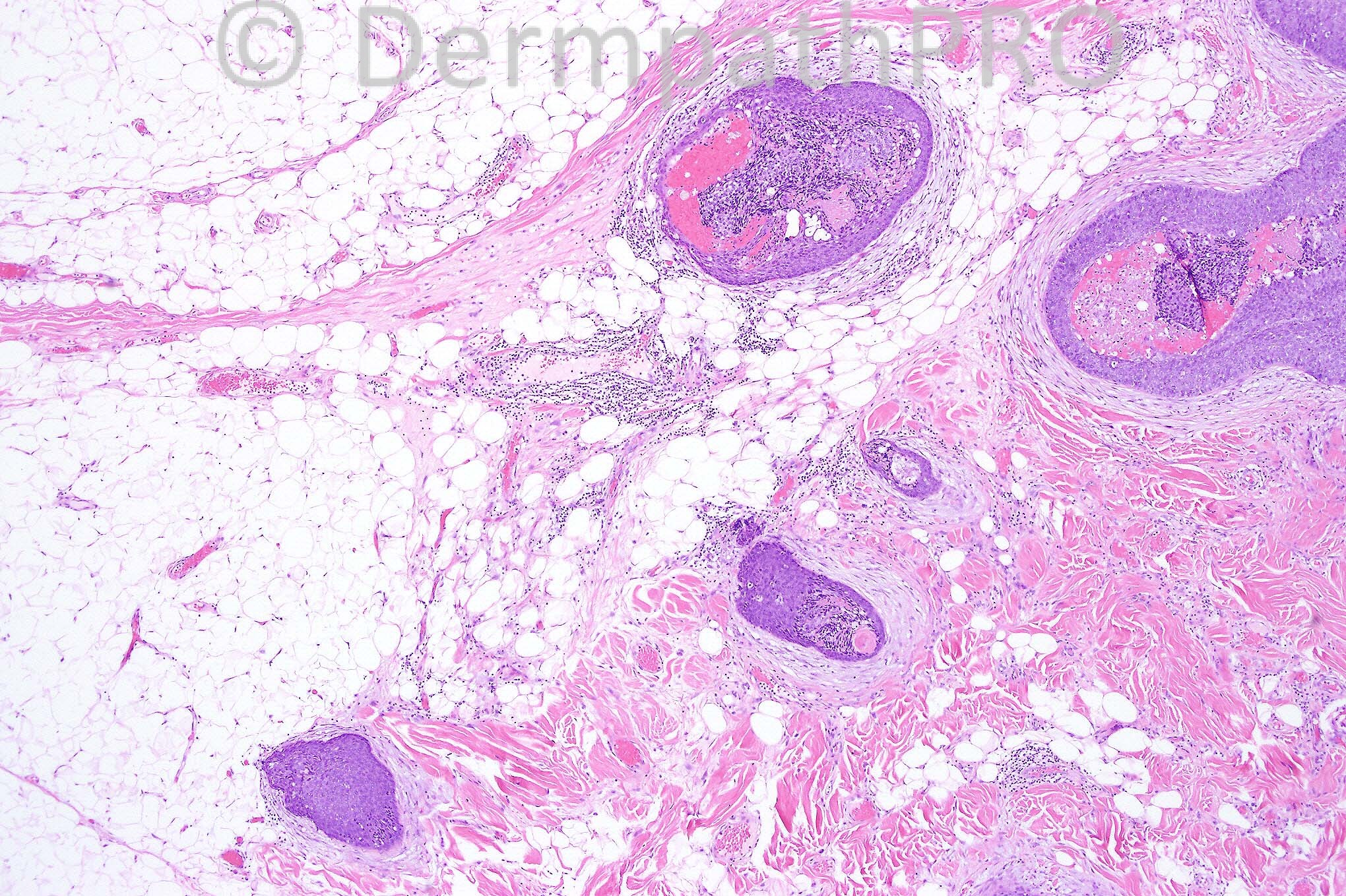
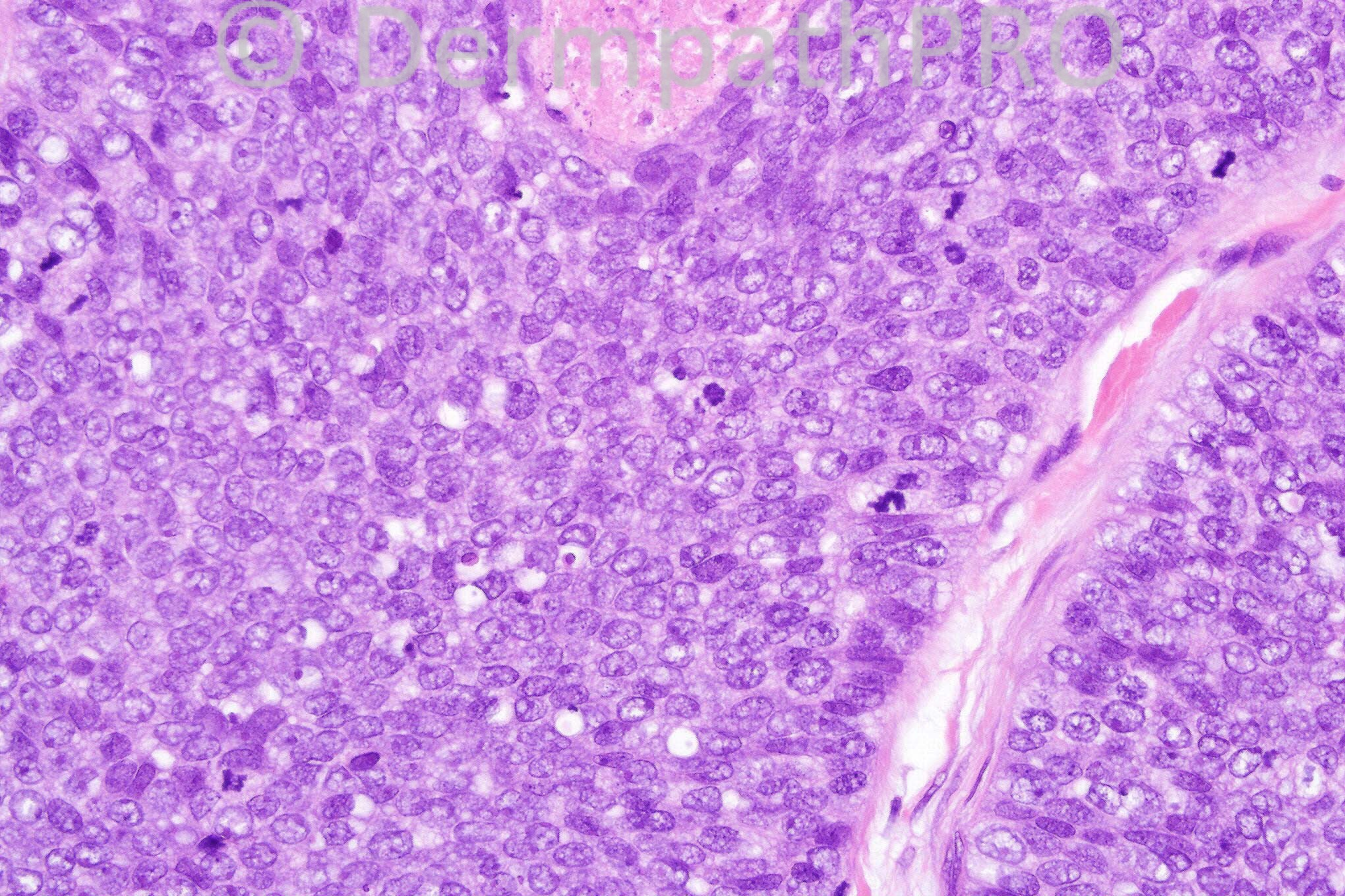
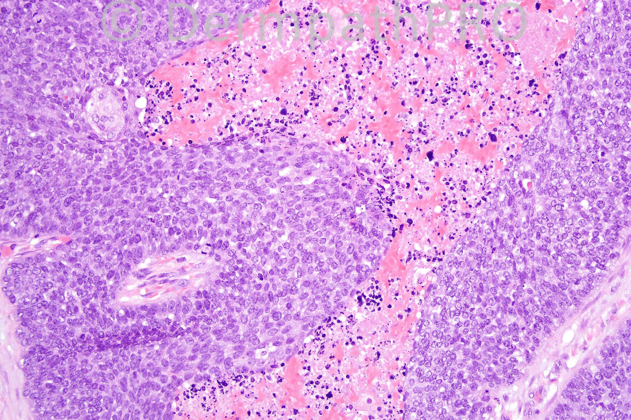
Male 84 years with a long-standing nodule on the scalp and recent increase in size.


  Report Record

User Feedback


error!! these are the photos of tommorow

Share this comment


Link to comment
share_externally

Guest Robledo F. Rocha

Posted

The images suggest pilomatrical carcinoma. There are interconnecting islands of basaloid cells with numerous mitoses. Shadow cells and nuclear debris are present in the center of the basaloid islands.

Share this comment


Link to comment
share_externally

Guest Guillermo Solis

Posted

Porocarcinoma with some areas of "comedo" necrosis

Share this comment


Link to comment
share_externally

Sasi Attili

Posted

Well, given the perineural invasion and the abnormal mitoses, this is a malignant adnexal neoplasm. I agree that the eosinophilic areas look more like comedo necrosis rather than shadow/ ghost cells. Similarly true trichilemmal keratinisation is not evident in these fields (considered malignant pilar tumour). Given the suggestion of ductal differentiation in the figure on the bottom left, I would favour a Porocarcinoma and confirm with CEA/ EMA.

Share this comment


Link to comment
share_externally

Arif Usmani

Posted

I agree with malignant adnexal neoplasm. A neoplasm that is perhaps arising in a nevus sebaceus given the long-standing history of a nodule with recent increase in size. The architecture reminds me of (malignant) dermal duct tumor so porocarcinoma is in the differential however I also see a few vacuoles (or at least one) indenting the nucleus which may be a clue to sebaceous carcinoma. In addition some of the nests exhibit prominent peripheral palisading and that raises a possibility of trichoblastic carcinoma (BCC variant?).

Share this comment


Link to comment
share_externally

Arif Usmani

Posted

Carcinomatous change in a pre-existing cylindroma is also a possibility which goes along with history.

Share this comment


Link to comment
share_externally

Sasi Attili

Posted

[quote name='Arif Usmani' timestamp='1354116948']
I agree with malignant adnexal neoplasm. A neoplasm that is perhaps arising in a nevus sebaceus given the long-standing history of a nodule with recent increase in size. The architecture reminds me of (malignant) dermal duct tumor so porocarcinoma is in the differential however I also see a few vacuoles (or at least one) indenting the nucleus which may be a clue to sebaceous carcinoma. In addition some of the nests exhibit prominent peripheral palisading and that raises a possibility of trichoblastic carcinoma (BCC variant?).
[/quote]
Agree with Seb carcinoma differential. However I am not able to convince myself of nuclear indentation

Share this comment


Link to comment
share_externally

Arif Usmani

Posted

Sasi as I said its very focal (fig 3) however can you appreciated jigsaw puzzling in the right mid aspect of fig-1. It may be a distraction.

Share this comment


Link to comment
share_externally

pilomatrix carcinoma > Porocarcinoma

Share this comment


Link to comment
share_externally

Dr. Mona Abdel-Halim

Posted

The appearance of the basaloid cells being attached to each other like a syncitium makes me favoring pilomatrical carcinoma.

Share this comment


Link to comment
share_externally

Dr. Phillip McKee

Posted

A difficult case made more so by only having 4 images. We all agree that it is malignant which is the most important thing. There is no real evidence of ductal differentiation although I can see why some of you might have thought that there was. The real problem is that I had the whole lesion to look at while you only have the images shown.. I agree with Mona and my diagnosis was pilomatrical carcinoma. There is true ghost cell change but unfortunately one cannot make it out in Fig 1.

Share this comment


Link to comment
share_externally


×
×
  • Create New...