Jump to content
In this section we have spot diagnoses posted on a daily basis since June 2010, now over 4000! You can review the archived cases and read the suggested diagnoses by users and the final comment by the contributors.
Case are uploaded each week day by 10 am UK time with the correct diagnosis will generally be posted at 8 pm UK time. Why not view the most recent spot diagnosis and proffer a diagnosis?

Case Number : Case 603 - 1 Oct Posted By: Guest

Please read the clinical history and view the images by clicking on them before you proffer your diagnosis.
Submitted Date :
   (0 reviews)

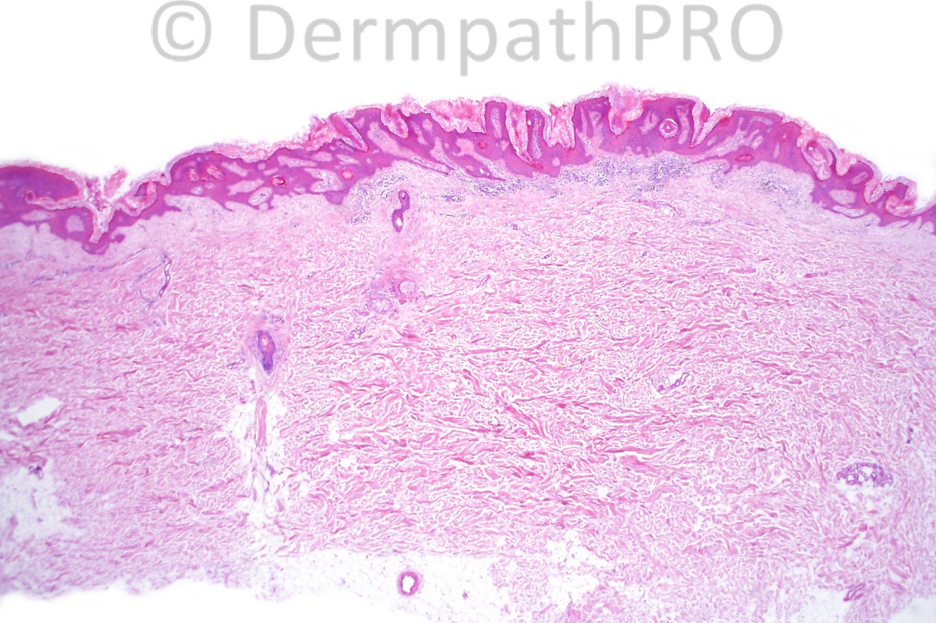
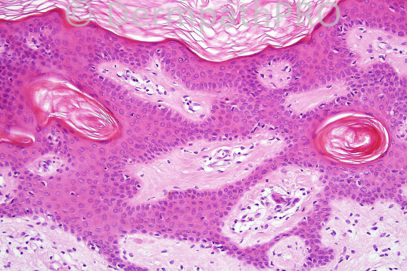
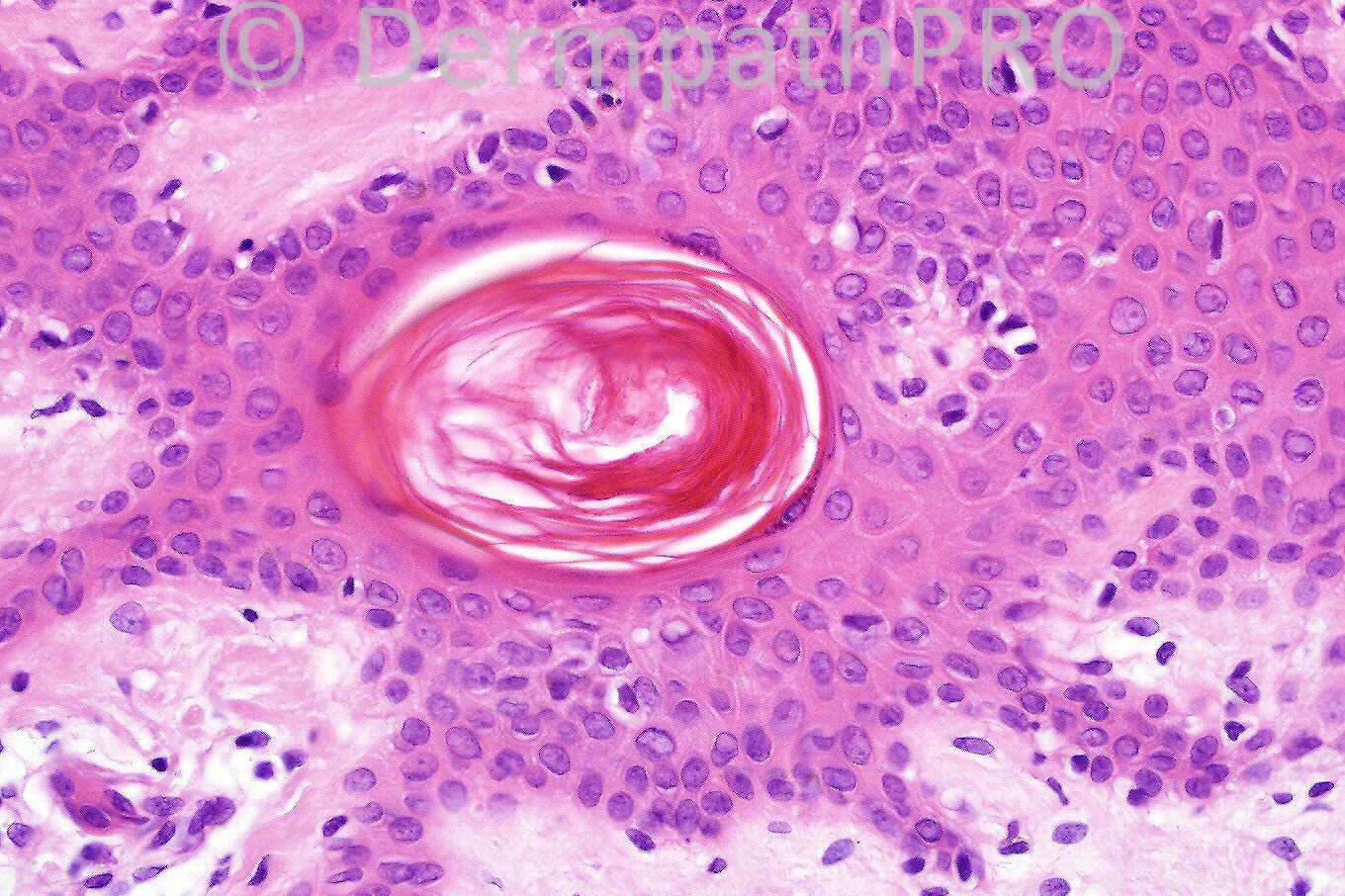
Male 6 years with a scaly, warty lesion on his arm.


  Report Record

User Feedback


Guest Amira Tawdy

Posted

Epidermal verruocus nevus or hyperkeratotic variant of seborreic keratosis, although there is some inward growth of the epidermis yet there is no koilocytes , no parakeratosis , no hypergranulosis to verify wart

Share this comment


Link to comment
share_externally

Guest Dr. Francisco Vílchez

Posted

Agree. Epidermal nevus.

Share this comment


Link to comment
share_externally

Hanan Vaknine MD

Posted

Epidermal nevus

Share this comment


Link to comment
share_externally

Guest Guillermo Solis

Posted

Epidermal nevus

Share this comment


Link to comment
share_externally

Guest Juan Carlos Garcés, Ecuador

Posted

Epidermal nevus

Share this comment


Link to comment
share_externally

Guest Dr Wilber J. Martínez

Posted

Epidermal nevus

Share this comment


Link to comment
share_externally

Dr. Mona Abdel-Halim

Posted

Epidermal Nevus

Share this comment


Link to comment
share_externally

Guest Dr. Shashi Baksh

Posted

Epidermal nevus

Share this comment


Link to comment
share_externally

Common or seb K-type of epidermal naevus.

Share this comment


Link to comment
share_externally

Dr. Phillip McKee

Posted

Epidermal nevus is correct. The age of the patient makes a diagnosis of seborrheic keratosis untenable.

Share this comment


Link to comment
share_externally

Guest Amr Gohar

Posted

[font="Times New Roman"][color="#000000"] [/color][/font]
[font="Times New Roman"][color="#000000"]Laminated hyperkeratosis – acanthosis – papillomatosis, the pattern that is usually seen in [b]verrucous epidermal naevus. [/b]It may resemble seborrhoeic keratosis at times.[b] Clinicopathological correlation[/b] is vital as always. [color=black]Almost all epidermal naevi follow the pattern of lines documented by Blaschko from drawings of epidermal naevi. The pattern is attributed to the lines of migration and proliferation of epidermal cells during embryogenesis. The lines do not correspond to any known nervous, vascular or lymphatic structures. The bands of abnormal skin represent clones of cells carrying a mutation in a gene expressed in the skin. Mosaicism impact on the phenotype relates to the proportion of cells that harbour the mutation and their distribution. Mosaic describes an art form in which pictures are produced by joining together tiny pieces of different coloured stone or glass. The pattern may vary according to cell type and timing of mosaicism. In the skin, [b]genetic mosaicism classically appears as Blaschko's lines[/b].[/color] Ep[b]idermolytic and non-epidermolytic verrucous epidermal naevi must be distinguished histopathologically[/b]. Patients with non-[font=courier new,courier,monospace]epidermolytic[/font][size="3"][font="Calibri"][color=black][font=courier new,courier,monospace] [/font][font=courier new,courier,monospace]epidermal naevi can be counselled that these are sporadic lesions and are not passed on as a generalized skin condition. Patients with epidermolytic verrucous epidermal naevi are at risk of parenting a child with bullous ichthyosiform erythroderma (BIE). If the patient is a child the carers should probably be informed of this risk, and of the possibility of first-trimester antenatal diagnosis.[/font][/color][/font][/size][/color][/font]

Share this comment


Link to comment
share_externally


×
×
  • Create New...