Jump to content
In this section we have spot diagnoses posted on a daily basis since June 2010, now over 4000! You can review the archived cases and read the suggested diagnoses by users and the final comment by the contributors.
Case are uploaded each week day by 10 am UK time with the correct diagnosis will generally be posted at 8 pm UK time. Why not view the most recent spot diagnosis and proffer a diagnosis?

Case Number : Case 604 - 2 Oct Posted By: Guest

Please read the clinical history and view the images by clicking on them before you proffer your diagnosis.
Submitted Date :
   (0 reviews)

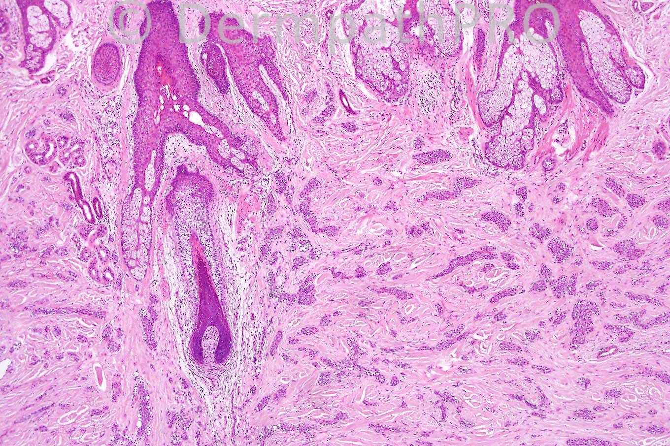
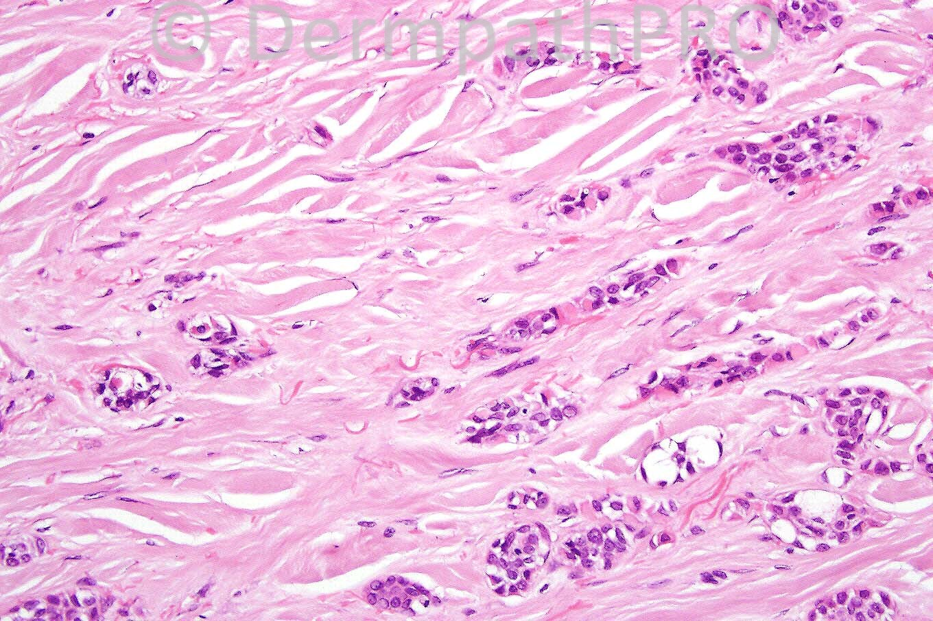
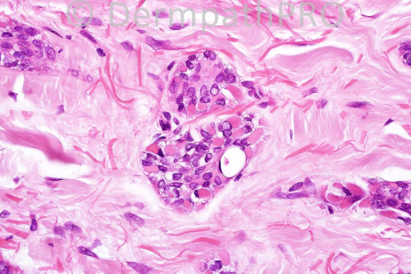
Female 62 years with a scaly indurated plaque on her face.


  Report Record

User Feedback


Hanan Vaknine MD

Posted

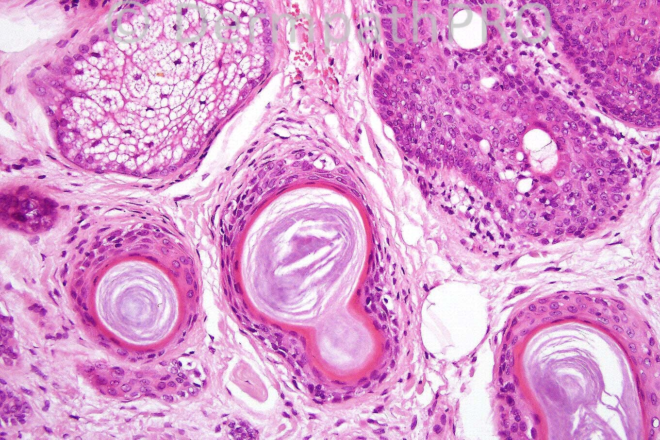
My first impression was of infiltrative BCC with signet ring formation (namely rhabdoid-signet ring basal cell carcinoma), however the keratin cyst and the vague glandular differentiation are more suggestive of Microcystic adnexal carcinoma.
Metastatic breast carcinoma (lobular with signet ring formation) should also be included in the differential diagnosis although less likely in this case

Share this comment


Link to comment
share_externally

Sasi Attili

Posted

My first impression on the images on top is a metastatic breast ca. However the bottom right horn cysts are slightly puzzling in this context. Not sure if it is a red herring. MAC is a possibility, but to low power is more in keeping with an infiltrative breast Ca.

Share this comment


Link to comment
share_externally

Guest Dr. Shashi Baksh

Posted

I agree my immediate impression based on the first three images was metastatic mammary carcinoma, but in light of the horn cysts I favor microcystic adnexal carcinoma.

Share this comment


Link to comment
share_externally

Guest Dr. Francisco Vílchez

Posted

I agree with Marcela and Shashi. Microcystic adnexal carcinoma.

Share this comment


Link to comment
share_externally

Guest Juan Carlos Garcés, Ecuador

Posted

MAC

Share this comment


Link to comment
share_externally

Guest Dr Wilber J. Martínez

Posted

[color=#000000][font=arial, sans-serif][size=3]Microcystic adnexal carcinoma[/size][/font][/color]

Share this comment


Link to comment
share_externally

Dr. Phillip McKee

Posted

I completely agree with the majority "vote". This is a classic MAC. The keratocysts in the 4th figure help make the diagnosis. Metastatic carcinoma with desmoplasia is a reasonable differential diagnosis. I am not keen on BCC. The examples of BCC with ductal differential that I have encountered have all been nodular variants with classic BCC features

Share this comment


Link to comment
share_externally


×
×
  • Create New...