Jump to content
In this section we have spot diagnoses posted on a daily basis since June 2010, now over 4000! You can review the archived cases and read the suggested diagnoses by users and the final comment by the contributors.
Case are uploaded each week day by 10 am UK time with the correct diagnosis will generally be posted at 8 pm UK time. Why not view the most recent spot diagnosis and proffer a diagnosis?

Case Number : Case 589 - 11 Sept Posted By: Guest

Please read the clinical history and view the images by clicking on them before you proffer your diagnosis.
Submitted Date :
   (0 reviews)

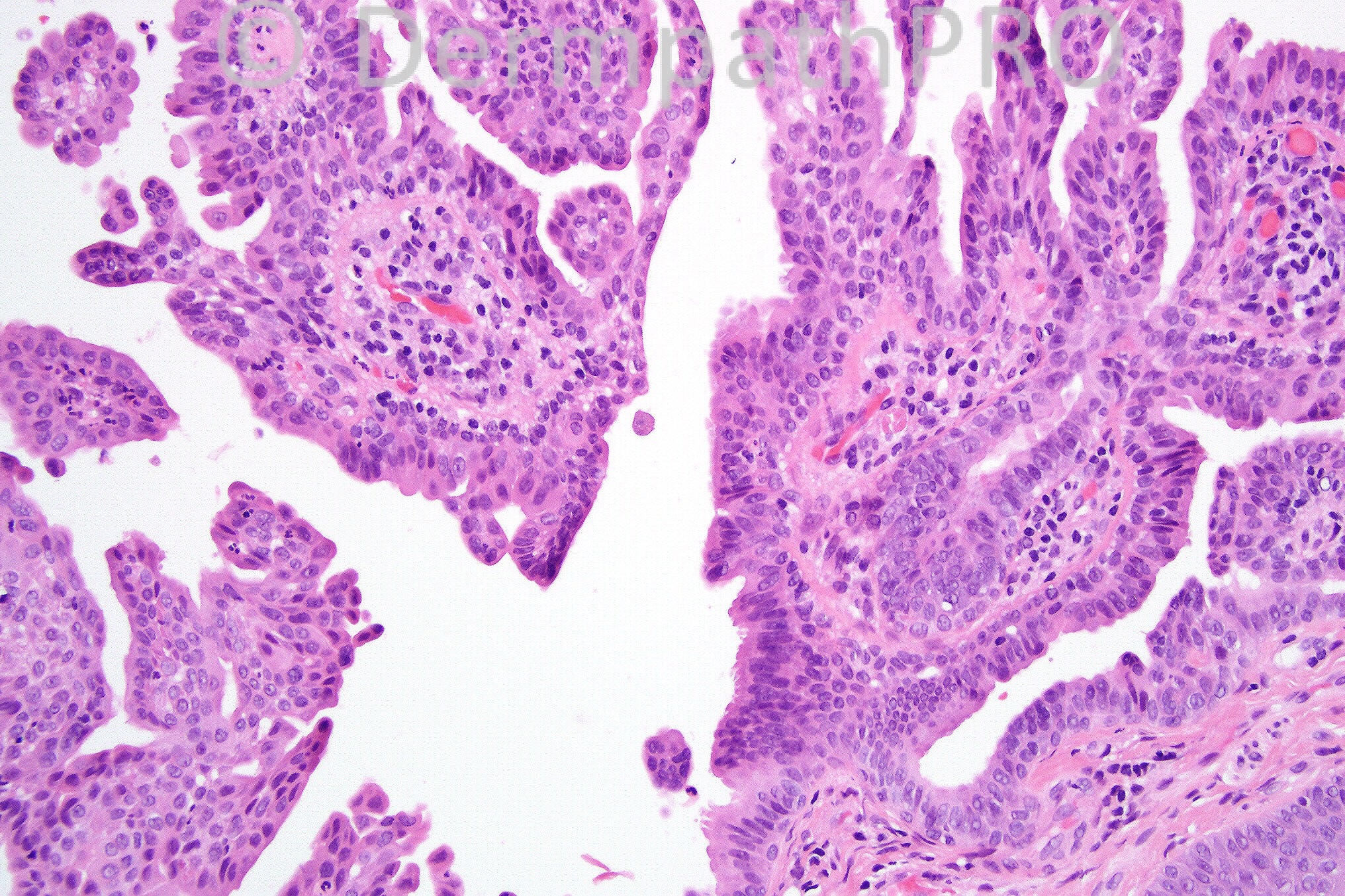
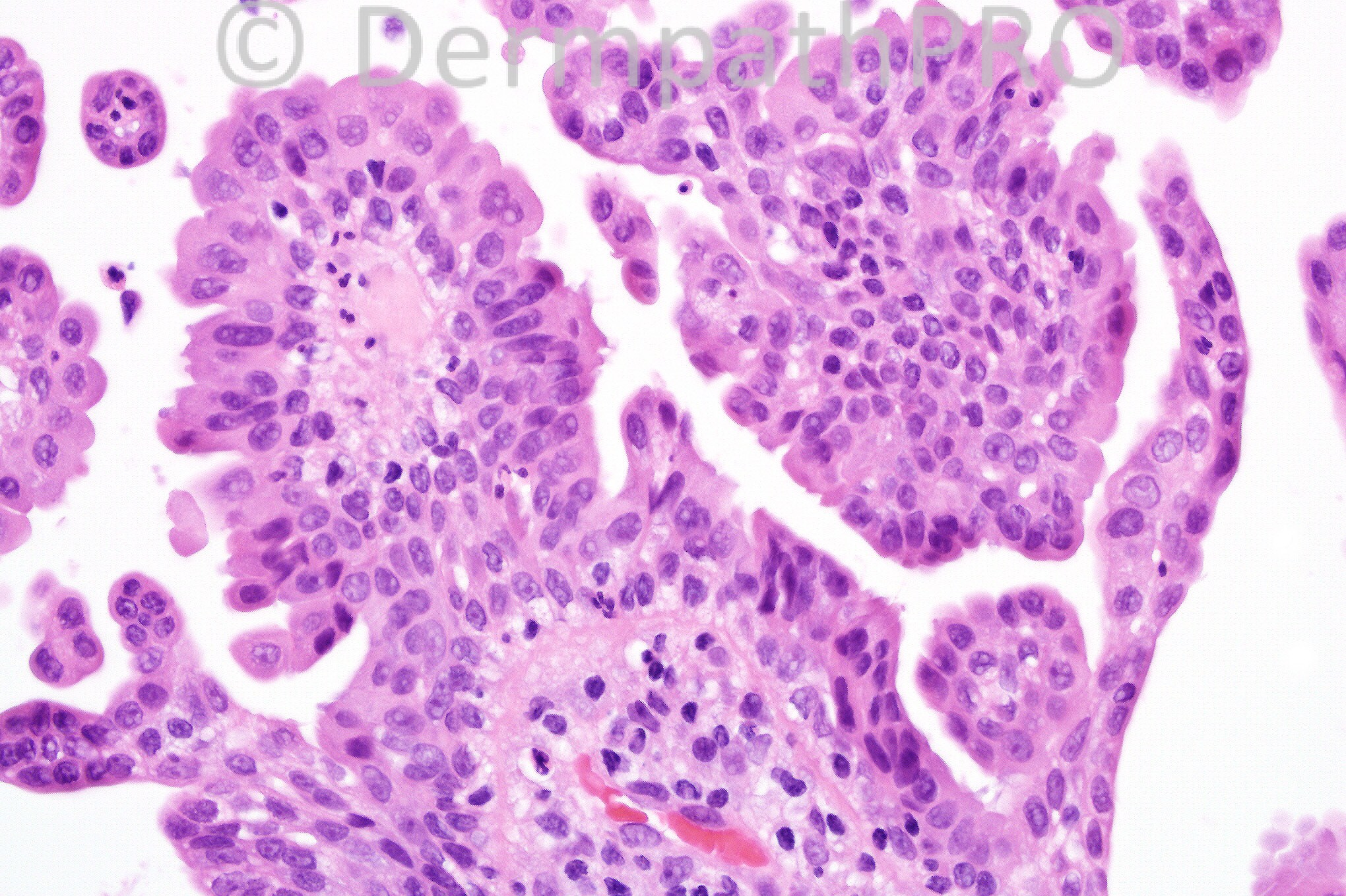
Female 43 years with a bluish lesion on the cheek. 


  Report Record

User Feedback


Sasi Attili

Posted

Apocrine cystadenoma, Syringocystadenoma papilliferum like...

Share this comment


Link to comment
share_externally

Guest Dr. Francisco Vílchez

Posted

It seems a papillary hidradenoma, though it usually appears in vulva...

Share this comment


Link to comment
share_externally

Favour syringocystadenoma papilliferum. Just wondered, would there be any management implication if this is an apocrine cystadenoma?

Share this comment


Link to comment
share_externally

Dr. Mona Abdel-Halim

Posted

Apocrine papillary cystadenoma,,, marvellous

Share this comment


Link to comment
share_externally

Guest Amr Gohar

Posted

[color="#000000"]Bluish hue – site – dermal cyst- question to be answered, is it [/color][color=#231F20]apocrine hidrocystoma, apocrine cystadenoma or [/color][color="#000000"]eccrine hidrocystoma? Because there is t[/color][color=#231F20]rue papillary projections (containing a fibrous core) this is [b]apocrine cystadenoma [/b](in apocrine hidrocystoma, the papillary projections are solid buds of secretory cells). Eccrine hidrocystoma often lies in [/color][color="#000000"]often in close proximity to eccrine glands with no evidence of decapitation secretion [/color][color=#231F20]n the cells of the[/color] [color=#231F20]inner layer.[/color]

Share this comment


Link to comment
share_externally

Guest Amr Gohar

Posted

Corrigenda
*there are
*secretion in

Share this comment


Link to comment
share_externally

Guest Amr Gohar

Posted

[color="#000000"]Bluish hue – site – dermal cyst- question to be answered, is it [/color][color=#231F20]apocrine hidrocystoma, apocrine cystadenoma or [/color][color="#000000"]eccrine hidrocystoma? Because there are t[/color][color=#231F20]rue papillary projections (containing a fibrous core) this is [b]apocrine cystadenoma [/b](in apocrine hidrocystoma, the papillary projections are solid buds of secretory cells). Eccrine hidrocystoma often lies [/color][color="#000000"]in close proximity to eccrine glands with no evidence of decapitation secretion i[/color][color=#231F20]n the cells of the[/color] [color=#231F20]inner layer.[/color]

Share this comment


Link to comment
share_externally

Hanan Vaknine MD

Posted

Apocrine papillary cystadenoma

Share this comment


Link to comment
share_externally

Dr. Phillip McKee

Posted

I don't suppose that it matters too much which label you apply! From my perspective, it is best regarded as an apocrine papillary cystadenoma. I agree that on the vulva you would call it a hidradenoma papilliferum. The fibrovascular cores seen in Figs 3 and 4 confirm that they are true papillae. Sometimes there may be an association with mevus sebaceus. Obviously, if this were part of the latter, there would be potential treatment implications because of cosmetic reasons and the inevitable development of other tumors including albeit rarely, carcinoma.

Share this comment


Link to comment
share_externally

Guest Marwa Fawzy

Posted

Apocrine papillary cystadenoma

Share this comment


Link to comment
share_externally


×
×
  • Create New...