Jump to content
In this section we have spot diagnoses posted on a daily basis since June 2010, now over 4000! You can review the archived cases and read the suggested diagnoses by users and the final comment by the contributors.
Case are uploaded each week day by 10 am UK time with the correct diagnosis will generally be posted at 8 pm UK time. Why not view the most recent spot diagnosis and proffer a diagnosis?

Case Number : Case 595 - 19 Sept Posted By: Guest

Please read the clinical history and view the images by clicking on them before you proffer your diagnosis.
Submitted Date :
   (0 reviews)

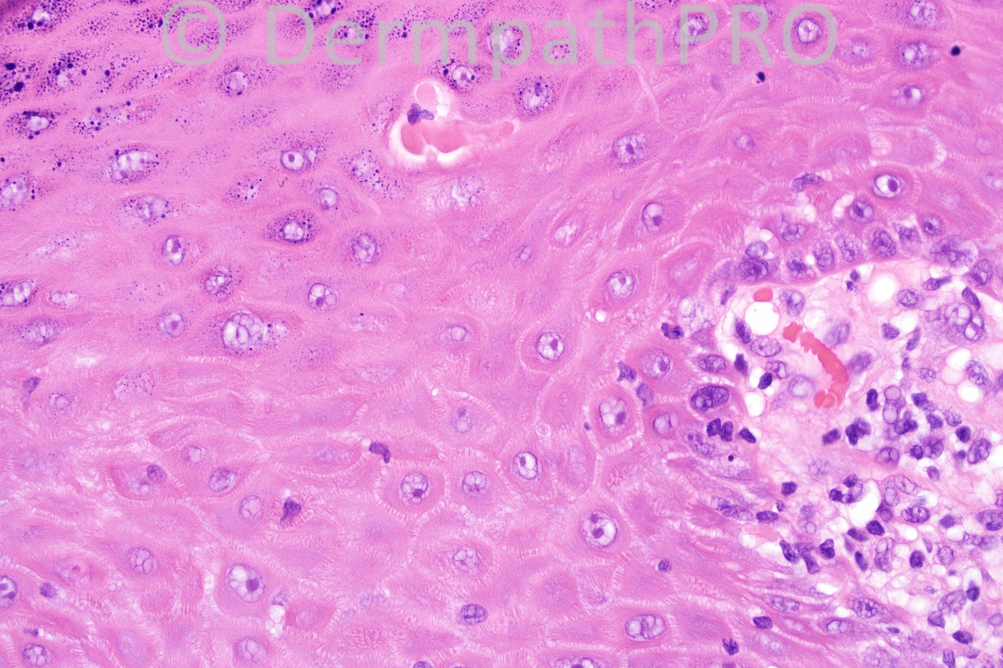
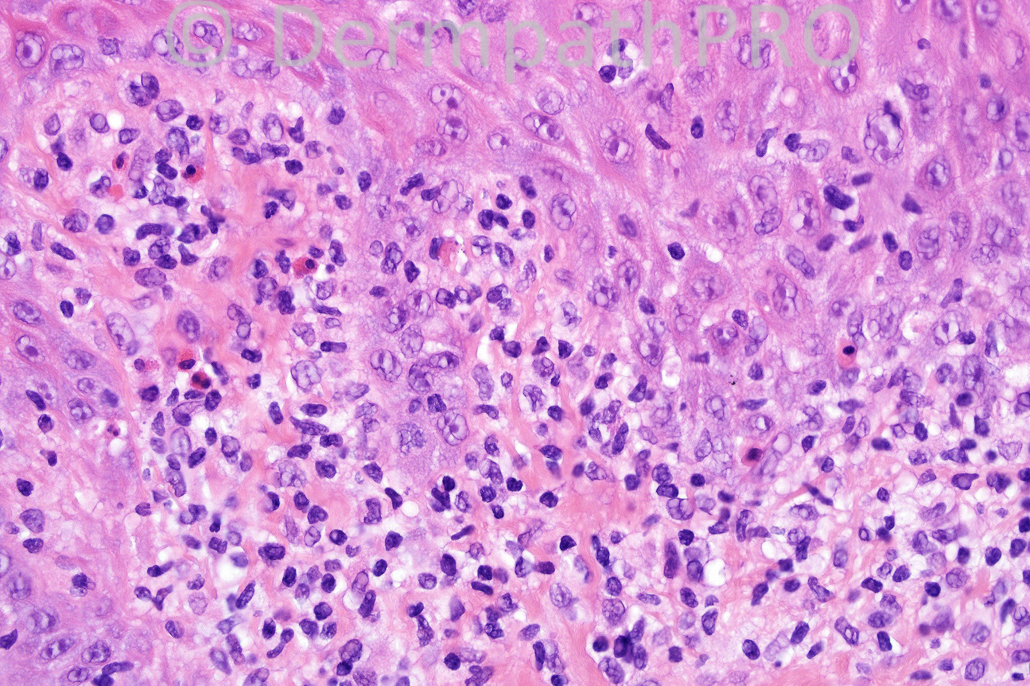
Male 49 years with an 1.0 com erythematous, scaly lesion on the face.


  Report Record

User Feedback


Concerned that this may represent patch stage MF. Would do Immunos.

Share this comment


Link to comment
share_externally

Guest Amira Tawdy

Posted

focal parakaeratosis, hyapergranulosis, interface dermatitis, band like lymphocytic infiltrate, few eosinphils I suggest drug induced lichenoid reaction

Share this comment


Link to comment
share_externally

Sasi Attili

Posted

agree with the histo description. Would add 'spongiosis' to the description and consider FDE and insect bite reaction in the differentials.

Share this comment


Link to comment
share_externally

Dr. Mona Abdel-Halim

Posted

Agree with the description, some plasma cells r also seen, with the clinical correlation (single lesion) will consider LPLK.

Share this comment


Link to comment
share_externally

lichenoid dermatitis with eos. Given clinical of solitary lesion: LPLK vs. solitary LP, perhaps FDE (if this is on lip.)

Share this comment


Link to comment
share_externally

LP-like keratosis vs LP-like drug reaction.

Share this comment


Link to comment
share_externally

Guest Guillermo Solis

Posted

Lichen planus Like keratosis

Share this comment


Link to comment
share_externally

Dr. Phillip McKee

Posted

Great. You have to take the clinical features into account otherwise you end up with a large differential diagnosis of lichenoid tissue reactions. The history of a solitary lesion is the give away. The only tenable diagnosis is a lichenoid keratosis. Lichenoid actinic keratosis is ruled out by the absence of atypia.

Share this comment


Link to comment
share_externally

Dr. Phillip McKee

Posted

I have posted several new comments on Facebook to announce some new features that will be available on the website. I hope that you like them. We have just started our test live video on melanoma variants. It will take a little time to iron out any problems but I am very excited about this new component.

Share this comment


Link to comment
share_externally


×
×
  • Create New...