Jump to content
In this section we have spot diagnoses posted on a daily basis since June 2010, now over 4000! You can review the archived cases and read the suggested diagnoses by users and the final comment by the contributors.
Case are uploaded each week day by 10 am UK time with the correct diagnosis will generally be posted at 8 pm UK time. Why not view the most recent spot diagnosis and proffer a diagnosis?

Case Number : Case 596 - 20 Sept Posted By: Guest

Please read the clinical history and view the images by clicking on them before you proffer your diagnosis.
Submitted Date :
   (0 reviews)

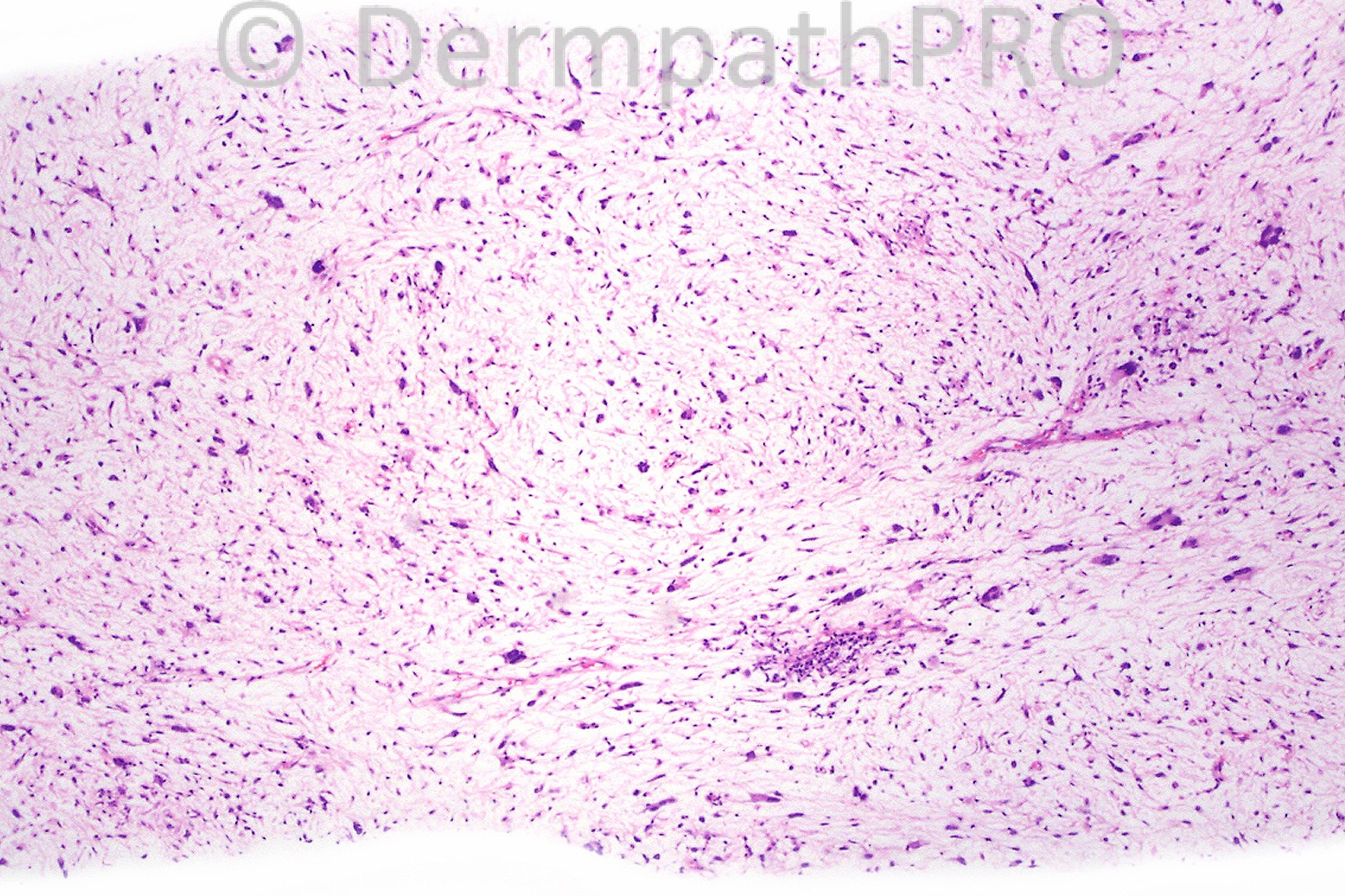
Male 60 years with a mass in the thigh.


  Report Record

User Feedback


Hanan Vaknine MD

Posted

[b]A core needle biopsy (therefore probably of a deep seated lesion) with myxoid stroma showing hyper-vascularity).[/b]
[b]No obvious lipoblasts seen, not even signet ring type[/b]
[b]No collagen bundles detected (which would suggest pleomorphic lipoma) [/b]
[b]Some floret like giant cells seen, at foci exhibiting atypia, devoid of mitotic figures[/b]
[b][u][b]My working diagnosis is myxoid liposarcoma with cellular atypia [/b][/u][/b]

Share this comment


Link to comment
share_externally

Sasi Attili

Posted

The lack of convincing lipoblasts and the crows feet type branching vessels to me suggest a myxofibrosarcoma. Looking forward to the actual diagnosis!

Share this comment


Link to comment
share_externally

Dr. Mona Abdel-Halim

Posted

I thought of myxofibrosarcoma.

Share this comment


Link to comment
share_externally

low grade fibromyxoid sarcoma / or hyalinizing spindle-cell tumor with giant rosettes

Share this comment


Link to comment
share_externally

Dr. Phillip McKee

Posted

These cases are always difficult without immunohistochemistry. The curvilinear vessels are a clue. The diagnosis proved to be myxofibrosarcoma. I do not like the pleomorphism and hyperchromatism. I would put it into the high grade category.

Share this comment


Link to comment
share_externally


×
×
  • Create New...