Jump to content
In this section we have spot diagnoses posted on a daily basis since June 2010, now over 4000! You can review the archived cases and read the suggested diagnoses by users and the final comment by the contributors.
Case are uploaded each week day by 10 am UK time with the correct diagnosis will generally be posted at 8 pm UK time. Why not view the most recent spot diagnosis and proffer a diagnosis?

Case Number : Case 728 - 1 Apr Posted By: Guest

Please read the clinical history and view the images by clicking on them before you proffer your diagnosis.
Submitted Date :
   (0 reviews)

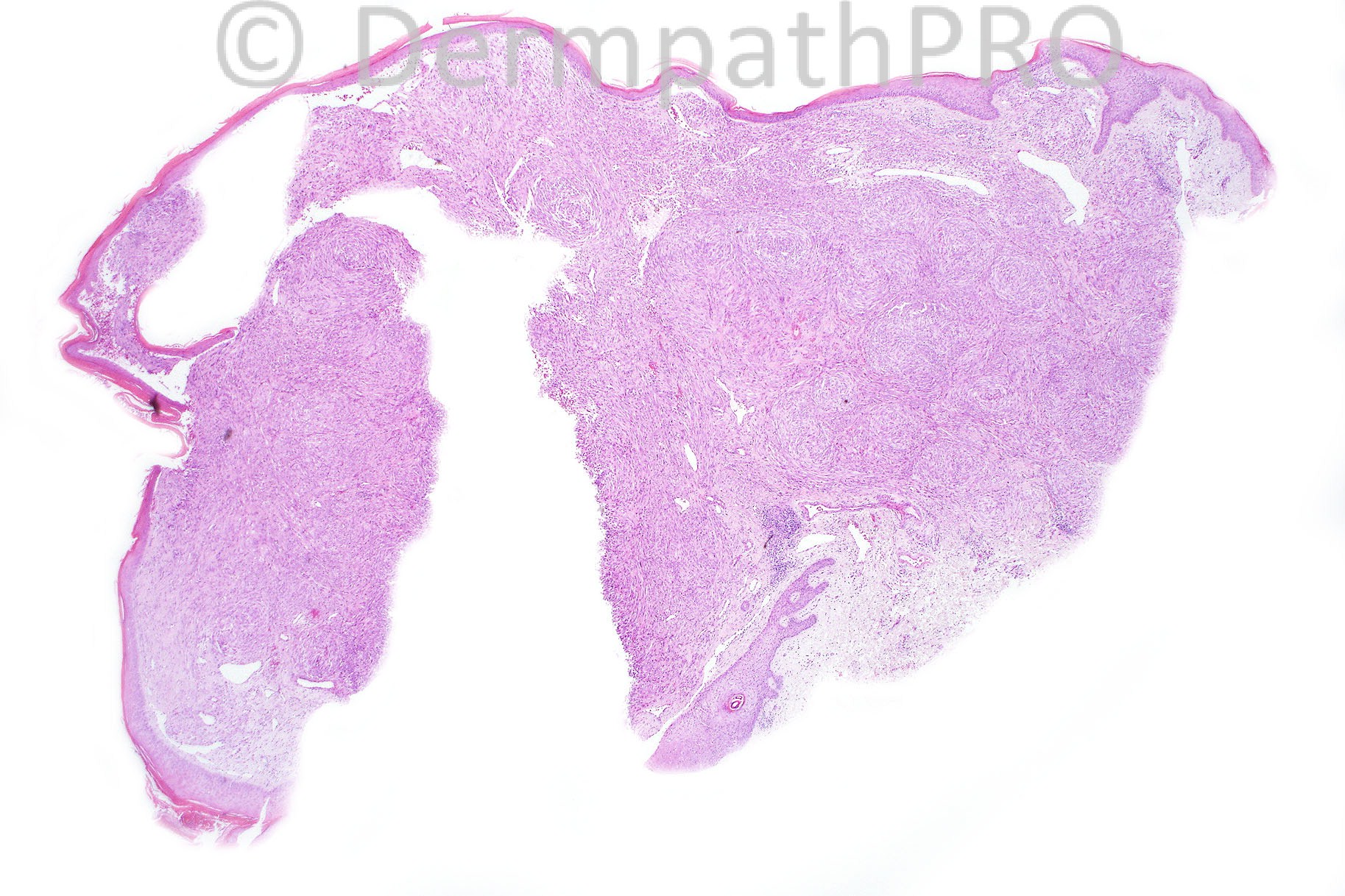
Lesion on the forearm of a 40 years old male.


  Report Record

User Feedback


Azza Esmat

Posted

Favor DFSP > cellular DF? for CD34/FXIIIa

Share this comment


Link to comment
share_externally

Dr. Mona Abdel-Halim

Posted

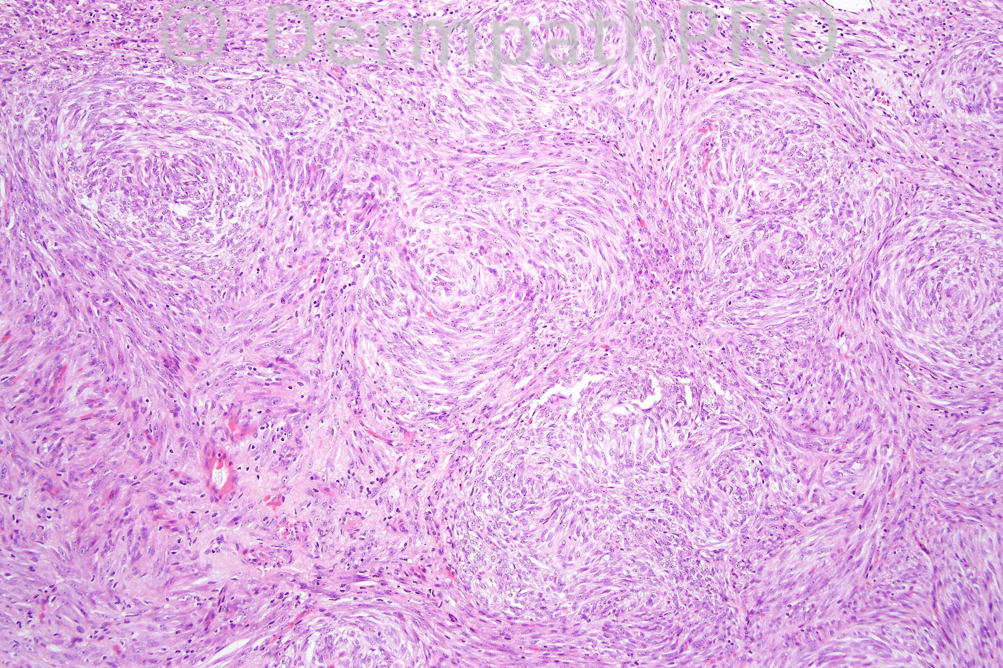
I percieved feathering in the second image and I can c foci of extravasated erythrocytes. Thought of nodular fasciitis.

Share this comment


Link to comment
share_externally

Eman El-Nabarawy

Posted

Cellular neurothecoma. DD perineuroma.

Share this comment


Link to comment
share_externally

Robledo F. Rocha

Posted

I favor cellular variant of nodular fasciitis. The lesion shows sharp circumscription and lacks nuclear atypia. Although there’s no prominent mucoid degeneration creating a tissue culture-like appearance, a myxoid stroma supports the sweeping fascicles of spindle fibroblast-like cells. Some scattered extravasated erythrocytes and ectatic vessels can also be appreciated.

Share this comment


Link to comment
share_externally

Dr. Phillip McKee

Posted

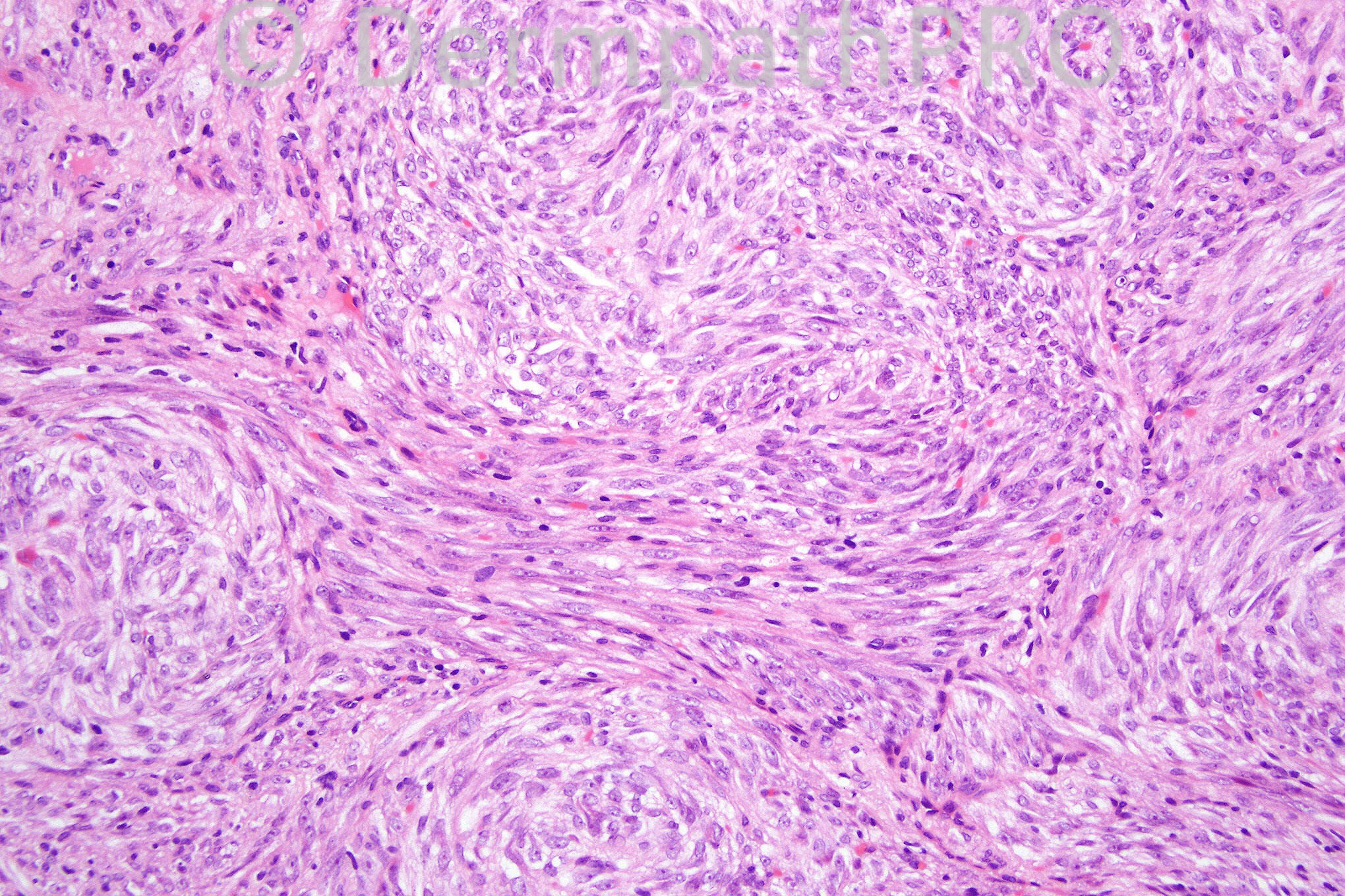
Spindle cell tumors in the skin are always a problem. This case (a gift from Richard Carr) was diagnosed as a superficial and solitary myofibroma. The biphasic pattern is seen to advantage in figure 4. It will be interesting to hear your views. Richard if you see this case, do you have any immunohistochemistry?

Share this comment


Link to comment
share_externally

Guest celso

Posted

[b]PERINEURIOMA, "meningioma-like" (the menigioma is not somehow a form of perineurium tumor?)[/b]

Share this comment


Link to comment
share_externally

Dr. Mona Abdel-Halim

Posted

I did not percieve the characteristic abundant esinophilic cytoplasm in the spindle cell fascicles which is characteristic of myofibroma at all!!! I think we need more explanation in this case !!!!

Share this comment


Link to comment
share_externally

Guest Romualdo C. L. Filho

Posted

I think the biphasic pattern created by whorls of bland spindle cells separated by vascular-rich areas with a vague pericytoma pattern is more in keeping with the diagnosis of myofibroma.

Share this comment


Link to comment
share_externally

Guest Jorge Gomila

Posted

Dermatofibrosarcoma protuberans de Darier Ferrand

Share this comment


Link to comment
share_externally

Dr. Richard Carr

Posted

THANK YOU PHILLIP - AS YOU KNOW SPINDLE CELL LESIONS ARE TRICKY FOR ME!!
This case was reported by a respected colleague as follows:
Curettings showing part of a bland dermal-based cellular lesion
comprising epithelioid and spindle cells in nested and fascicular
arrangement with storiform areas. Occasional mitotic figures and
background inflammation with vascularity highlighted by CD34.
Tumour cells are negative for S100, Melan-A, Desmin and CD34. Weak
non-specific staining with CD68. Features are of a benign mesenchymal
and tumour, dermatofibroma is one possibility although the fascicular
nature is unusual. SUPPLEMENTARY REPORT:
Additional immunostains show positivity for Smooth muscle actin
confirming a myofibroblastic origin, and this stain indirectly also
highlights a biphasic pattern of fascicular areas contrasting with more
haemangiopericytomatous areas. These features are supportive of a
diagnosis of benign myofibroma.
MY (DR CARR) THOUGHTS: I took a look at the lab record and the report does not
state but EMA and NKI (CD57) were done as I am sure with this morphology
both cellular neurothekeoma and perineurioma were considerations.
I can only assume they were negative. I pesonally would still
think about an epithelioid variant of dermatofibroma in this case - I have
looked at the section again (in my slide collection) and I do note quite a bit of
haemosiderin and a rather polypoid (slightly cross-cut) appearance. I am happy
to put it in to a benign myofibroblastic category.
Great case for exciting discussion!!

Share this comment


Link to comment
share_externally

Dr. Mona Abdel-Halim

Posted

Thanks Dr Carr for this nice case.

Share this comment


Link to comment
share_externally


×
×
  • Create New...