Jump to content
In this section we have spot diagnoses posted on a daily basis since June 2010, now over 4000! You can review the archived cases and read the suggested diagnoses by users and the final comment by the contributors.
Case are uploaded each week day by 10 am UK time with the correct diagnosis will generally be posted at 8 pm UK time. Why not view the most recent spot diagnosis and proffer a diagnosis?

Case Number : Case 730 - 3 Apr Posted By: Guest

Please read the clinical history and view the images by clicking on them before you proffer your diagnosis.
Submitted Date :
   (0 reviews)

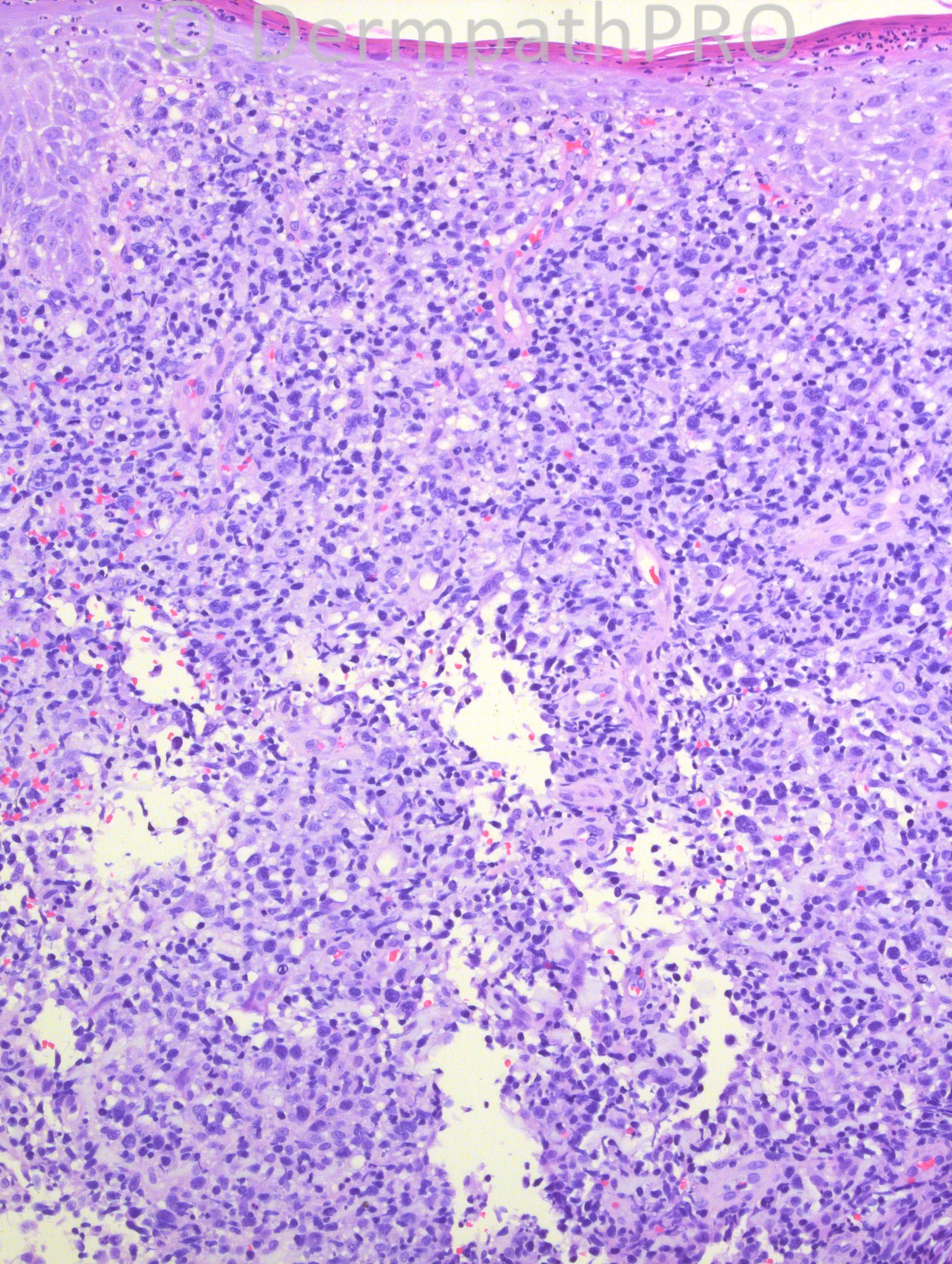
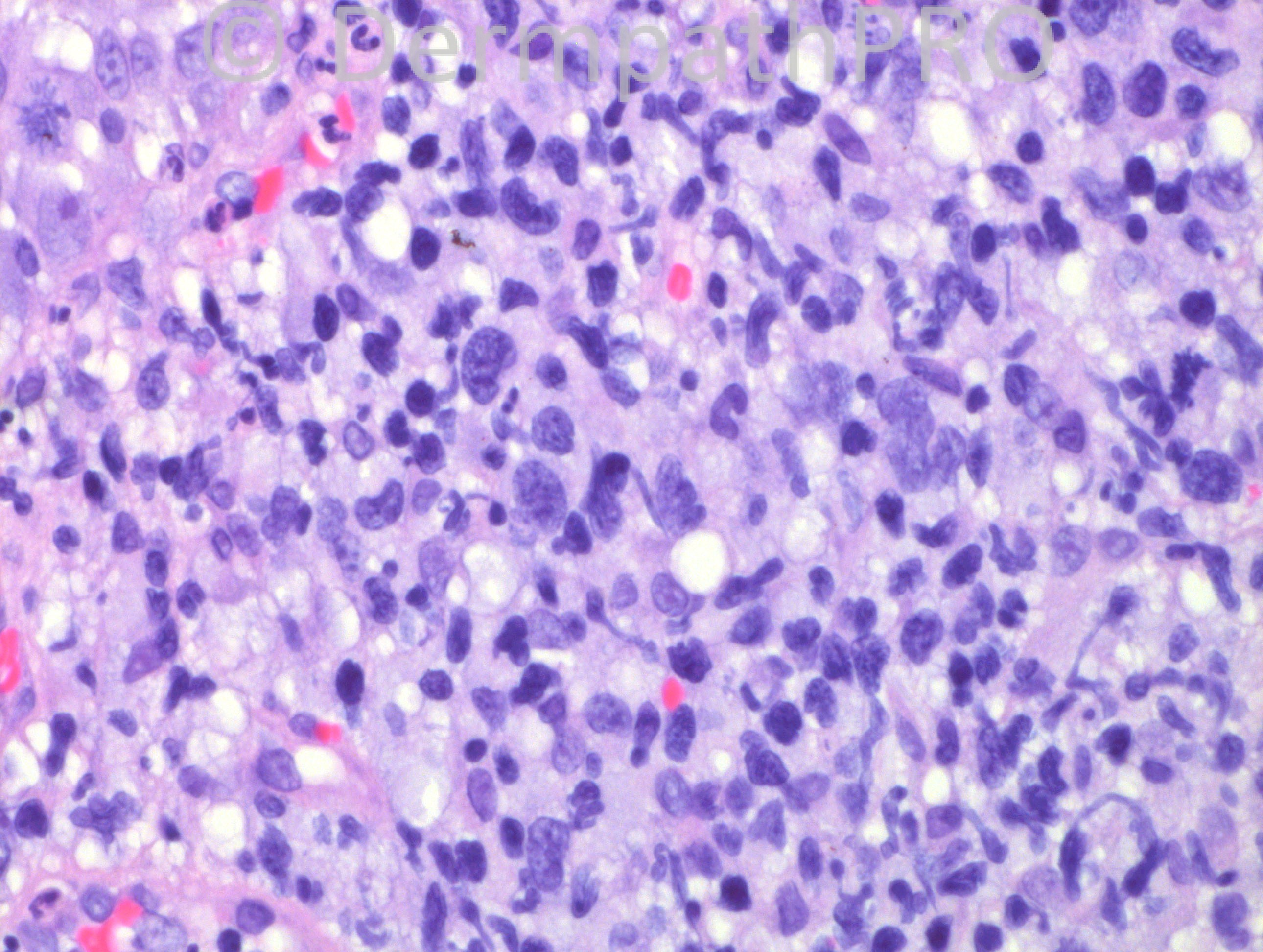
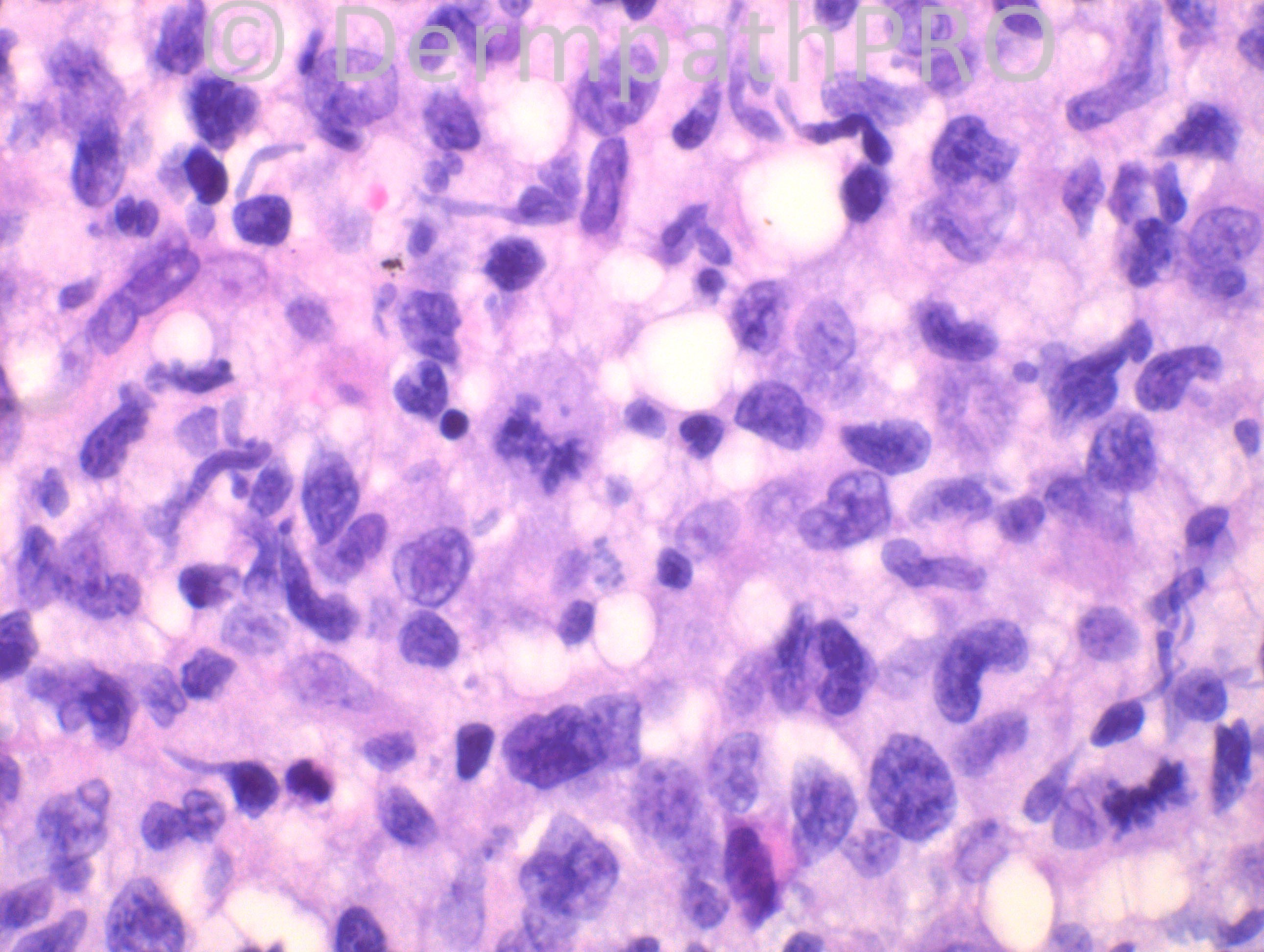
62 years-old male with a nodule on his right upper arm, 4 cm above elbow. Clinical differential diagnosis: BCE, squamous cell carcinoma, melanoma, metastatic carcinoma, lymphoma.

Case posted by Dr. Hafeez Diwan.


  Report Record

User Feedback


Guest Dr Engin Sezer

Posted

DDx including LCH or lymphomas (CD30 anaplastic vs. others)

Share this comment


Link to comment
share_externally

Sasi Attili

Posted

My first impression was a cutaneous CD30+ve ALCL. Obviously needs immunos to confirm and rule out other differentials mentioned!

Share this comment


Link to comment
share_externally

Guest Rodrigo Restrepo

Posted

LCL CD30+

Share this comment


Link to comment
share_externally

The intracytoplasmic vacuoles and extravasated RBCs made me think of vascular lineage of the lesion, I would consider epitheloid angiosarcoma in the DD, immunos are mandatory.

Share this comment


Link to comment
share_externally

Guest Bansal_

Posted

Favour cutaneous large cell lymphoma, likely CD30 positive.

Share this comment


Link to comment
share_externally

Robledo F. Rocha

Posted

Sheets of cohesive atypical cells infiltrating diffusely, at least, within the entire dermis. Neoplastic cells have large rounded or irregularly shaped nuclei with prominent nucleoli and abundant cytoplasm. Epidermotropic cells and imminent ulceration are other features.
I favor anaplastic large cell lymphoma. A panel of immunostains is mandatory to rule out mimickers. Clinical correlation is crucial in order to confirm if it is primary cutaneous or represent a secondary involvement of the skin by systemic disease.

Share this comment


Link to comment
share_externally

Guest Maria george

Posted

Sebaceous carcinoma, xanthogranuloma in the differential but I favor Large cell lymphoma .CD20, , CD 30 are a must.

Share this comment


Link to comment
share_externally

Dr. Mona Abdel-Halim

Posted

I can identify many large atypical cells with embryo shaped nuclei and many mitoses, so my first impression is anaplastic large TCL, will definitely go for Immunos: CD3 and CD30. There is no grenz zone, but CD20 to be done to complete the profile.

Share this comment


Link to comment
share_externally

Guest Jim Davie MD

Posted

[size=4]Tough one given our limited photo milieu. The epidermal involvement and diffuse non-cohesive infiltrate leans towards lymphoma/infectious pseudolymphoma. [/size][size=4]I agree the ?pseudovascular vs. vascular clefts and extensive vacuolation are of interest...doubt but ?angiosarc/liposarc. Is that granular dark pigment in the bottom two photos? [/size]
- Neoplastic lymphoma, rule out infectious (CD3,CD5,CD10,CD30, CD79a, PAS to start)
- Neoplastic non-lymphoma, poorly differentiated (CD31, S100, CAM5.2/LMW cytokeratin, HMW cytokeratin to start).

Share this comment


Link to comment
share_externally

Eman El-Nabarawy

Posted

Large Anaplastic TCL.

Share this comment


Link to comment
share_externally

Also suspect a C-ALCL. Thinking in Lymphoma and as a isolated lesion, there´s hardly other dif. diagnosis.

Share this comment


Link to comment
share_externally

Dr. Hafeez Diwan

Posted

Agreed. This was cutaneous ALCL. There are no other lesions. CD30 was diffusely positive and ALK-1 was negative.

Share this comment


Link to comment
share_externally


×
×
  • Create New...