Jump to content
In this section we have spot diagnoses posted on a daily basis since June 2010, now over 4000! You can review the archived cases and read the suggested diagnoses by users and the final comment by the contributors.
Case are uploaded each week day by 10 am UK time with the correct diagnosis will generally be posted at 8 pm UK time. Why not view the most recent spot diagnosis and proffer a diagnosis?

Case Number : Case 737 - 12 April Posted By: Guest

Please read the clinical history and view the images by clicking on them before you proffer your diagnosis.
Submitted Date :
   (0 reviews)

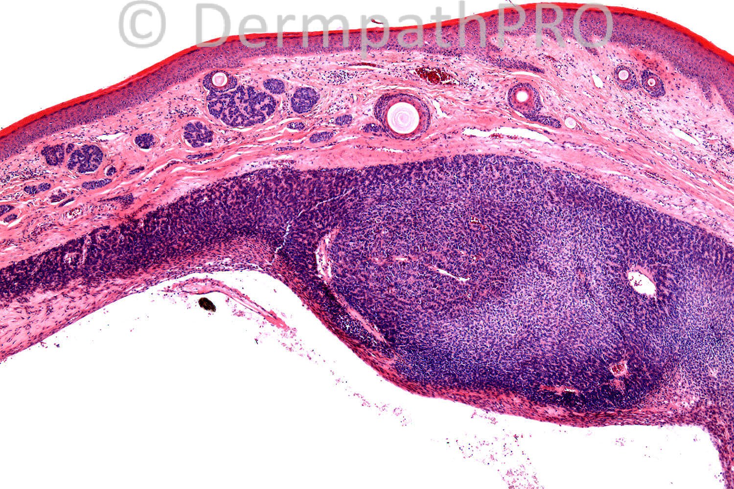
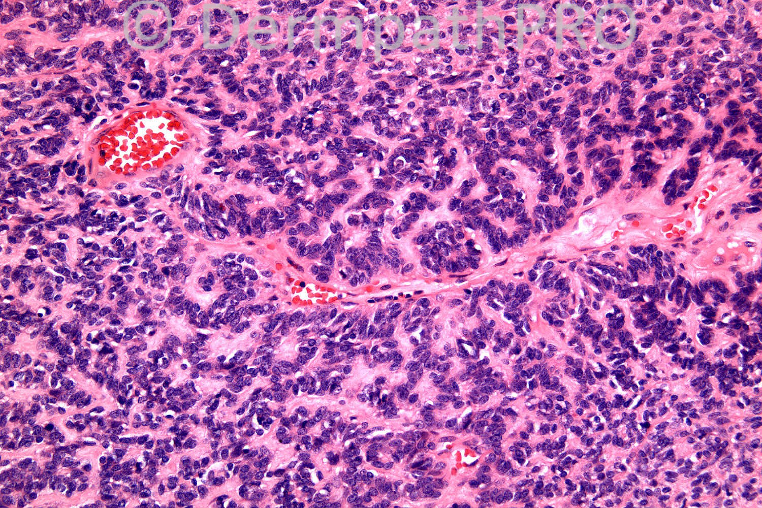
56 years-old male. Cyst on scalp for two years. Increased in size. ?Malignant.

Case posted by Dr. Richard Carr.


  Report Record

User Feedback


Eman El-Nabarawy

Posted

Sebaceoma.

Share this comment


Link to comment
share_externally

Guest Dr Engin Sezer

Posted

Trichoblastoma with sebaceous differentiation

Share this comment


Link to comment
share_externally

Guest Dr Engin Sezer

Posted

I like the rippled pattern in trichoblastoma:)

Share this comment


Link to comment
share_externally

Dr. Mona Abdel-Halim

Posted

I have two suggestions here: either this is a rippled pattern trichoblastoma with focal sebaceous differentiation (such sebaceous differentiation is common in this type of trichoblastoma). OR this is a collision lesion of trichoepithelioma in the superficial dermis and a sebaceoma lying deeper with a rippled pattern.

Share this comment


Link to comment
share_externally

Sasi Attili

Posted

[color=#1C2837][font=arial, verdana, tahoma, sans-serif][size=4] rippled pattern sebaceous trichoblastoma[/size][/font][/color]

Share this comment


Link to comment
share_externally

Eman El-Nabarawy

Posted

I think cystic changes are more in sebaceoma!!

Share this comment


Link to comment
share_externally

Guest Dr Engin Sezer

Posted

Let's perform CD20 to highlight Merkel cell colonization then:))

Share this comment


Link to comment
share_externally

Robledo F. Rocha

Posted

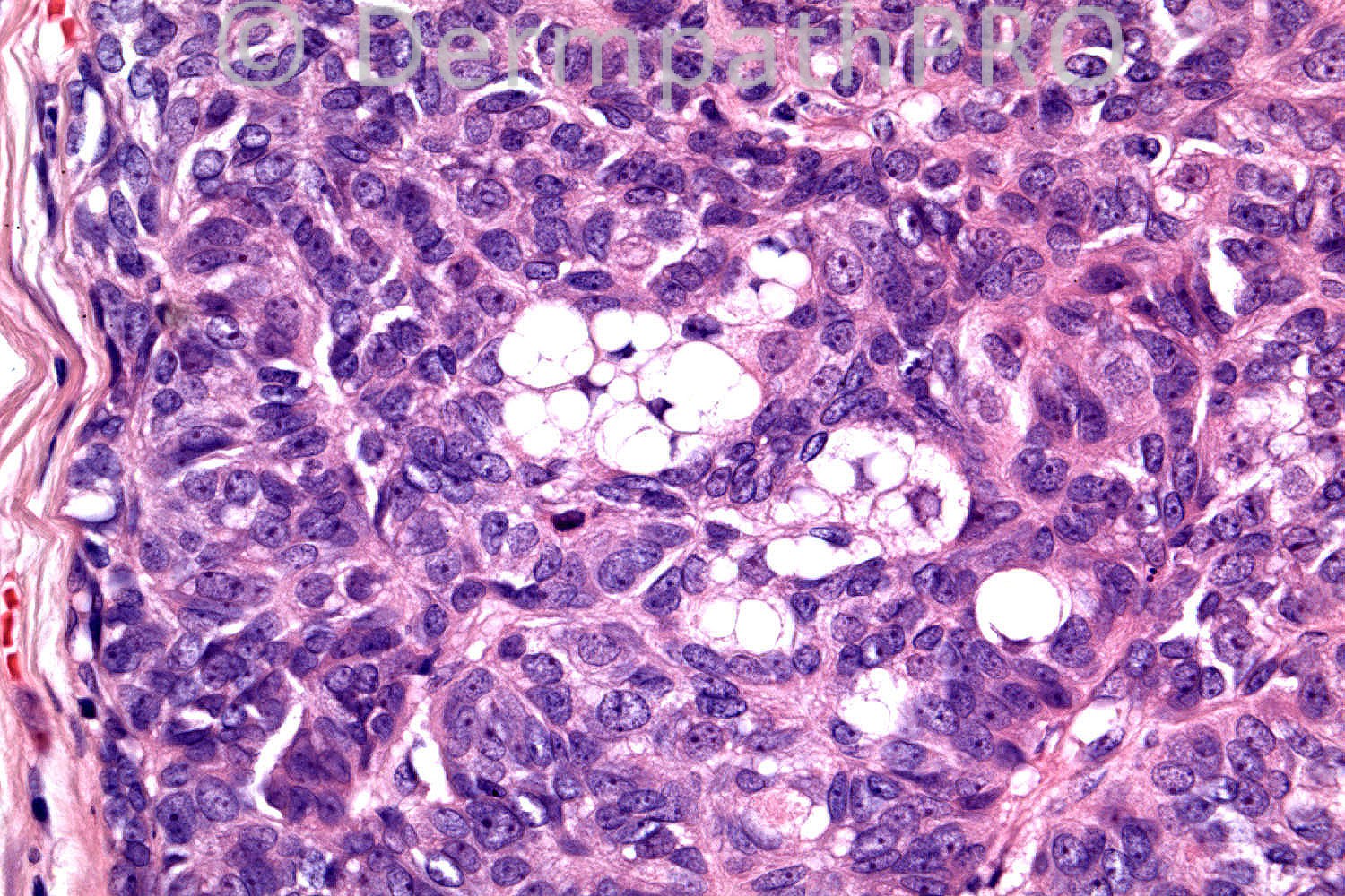
Solid-cystic tumor composed predominantly of small basaloid cells arranged in a rippled pattern. There are admixed scarce sebocytes and small cystic structures. The last seem to have an inner hyaline cuticular lining, what suggests that they are sebaceous ducts rather than keratocysts.
So, I think it is sebaceoma.

Share this comment


Link to comment
share_externally

Guest Dr Engin Sezer

Posted

My question is how one can explain the keratocysts in the upper dermis and relate those with sebaceoma? On the other hand, if CD20 was negative I'd definetely perform MLH-1, MSH-2,6 to rule out Muir-Torre..

Share this comment


Link to comment
share_externally

Dr. Mona Abdel-Halim

Posted

The question raised by Dr Engin is the reason behind me putting the rippled trichoblastoma with seb diff as a first possibility, however, if we think of a collision lesion, we might explain the keratocysts as trichoepithelioma ssociated with sebaceoma in the same lesion. Nice case!!!!!

Share this comment


Link to comment
share_externally

Guest Dr Engin Sezer

Posted

I completely agree, and I had already seen a case with trichoep in the upper dermis with cylindroma in the deeper dermis at St. John's. That's why I fell that demonstrating the Merkel cell colonization may be helpful to differentiate both (i.ie trichoblastoma vs. sebaceoma). Curious regarding the result though, very interesting case and arguement

Share this comment


Link to comment
share_externally

Robledo F. Rocha

Posted

[quote name='Dr. Mark Hurt' timestamp='1365770149']
Question for the panel: Do you regard these as potentially part of the Muir-Torre Syndrome, or do you regard them, as Ackerman did, as unrelated to that syndrome?
[/quote]
Latest data from the literature attest that sebaceoma, like sebaceous adenoma (the so-called “most specific marker”), have significantly high rates of mutations related to Muir-Torre syndrome, mainly if this tumor occurs outside of the head and neck region (see [url="http://www.ncbi.nlm.nih.gov/pmc/articles/PMC2805836/pdf/nihms158792.pdf"]http://www.ncbi.nlm....nihms158792.pdf[/url]).
Anyway, I think any sebaceous neoplasm must recommend screening for Muir-Torre syndrome.

Share this comment


Link to comment
share_externally

Robledo F. Rocha

Posted

[quote name='Dr Engin Sezer' timestamp='1365774039']
My question is how one can explain the keratocysts in the upper dermis and relate those with sebaceoma?
[/quote]
[color=#1C2837][font=arial, verdana, tahoma, sans-serif][size=4]As I wrote above, I think that those [/size][/font][/color][color=#1C2837][font=arial, verdana, tahoma, sans-serif][size=4]small cystic structures[/size][/font][/color][color=#1C2837][font=arial, verdana, tahoma, sans-serif][size=4] seem to have an inner hyaline cuticular lining, what drives me to interpret them as sebaceous ducts rather than keratocysts.[/size][/font][/color]

Share this comment


Link to comment
share_externally

Sebaceoma with a rippled pattern.

Share this comment


Link to comment
share_externally

Dr. Richard Carr

Posted

Great discussion. Clearly some of you have not already bought Dr Kazakov's book (Phillip did much of the photography). I will quote verbatim as follows: "A constant finding in sebaceous neoplasms dominated by organoid arrangements (labyrinthine/sinusoidal, rippled, petaloid, and carcinoid-like patterns) is the paucity of mature sebocytes. There are rare cases practically devoid of mature sebocytes, often requirig multiple sections to identify them. This often results in misinterpretation of such lesions as trichoblastomas, including so-called rippled pattern trichoblastoma. Ohata and Akerman, after critically reviewing published cases of the so-called rippled trichoblastomas, suggested that all these tumours, in fact, represent sebaceous neoplasms; the authors concur wtih this conclusion. In fact, the distinction between sebaceous lesions and genuine trichoblastoma, a benign follicular neoplasm with biphasic epithelial-mesenchymal follicular differentiation, is straightforward since sebaceous neoplasms always lack the typical follicular [papillary mesenchymal cell - RAC] stroma found in trichoblastomas."

Fig 3.47 and Fig 3.48 in Kazakov's book also highlights the infundibular keratocytes [present in my case] in a sebaceoma that in Fig 3.48 are so prominent they mimic trichadenoma. Fig 3.50 shows a case with prominent stromal hyalinsation [as was also seen in my case] and can give a resemblance to cylindroma or spiradenoma. Kazakov's book also mentions non-sebceous duct including apocrine ductal differentiation in some sebaceous associated with p63 positive myoepithelial cells.

Regarding the current case sorry Engin I did not do a CK20 for Merkel cells as I had diagnosed sebaceoma on H&E and supported by lack of BerEP4 (all our TBs have at lease focal moderate BerEP4 expression). p63 was diffusely expressed in the case. Incidentally the MMR staining was preserved and since this appeared as a solitary lesion on the head no further recommendation for genetic studies for Muir Torre was undertaken. I e-mailed Dmitry Kazakov who comments he is currently studying the MMR's in the variants of sebaceoma so we may get some news in due course in answer to Marks question which I assumed related to the rippled variant. Rember to consider the possibility of Muir Torre in all cases of sebaceous neoplasms particularly when multiple and for all lesions arising off the head. I normally comment in reports of sebaceous carcinomas of the eyelid and other solitary sebaceous lesions presenting on the head in the elderly that the association with Muir Torre is minimal.

Thank you all for another great discussion and do enjoy your weekends.

Share this comment


Link to comment
share_externally

Guest Romualdo C. L. Filho

Posted

Rippled pattern sebaceous trichoblastoma.

Share this comment


Link to comment
share_externally

Dr. Mona Abdel-Halim

Posted

Great case, amazing discussion.... Will buy Kasakov book immeadiately :-) Thanks Dr Carr,,,

Share this comment


Link to comment
share_externally

Robledo F. Rocha

Posted

Me too, Mona!

Share this comment


Link to comment
share_externally

Guest Peter Talarcik

Posted

:) rather obsolete discussion, it is clearly rippled pattern sebaceoma

Share this comment


Link to comment
share_externally


×
×
  • Create New...