Jump to content
In this section we have spot diagnoses posted on a daily basis since June 2010, now over 4000! You can review the archived cases and read the suggested diagnoses by users and the final comment by the contributors.
Case are uploaded each week day by 10 am UK time with the correct diagnosis will generally be posted at 8 pm UK time. Why not view the most recent spot diagnosis and proffer a diagnosis?

Case Number : Case 738 - 15 Apr Posted By: Guest

Please read the clinical history and view the images by clicking on them before you proffer your diagnosis.
Submitted Date :
   (0 reviews)

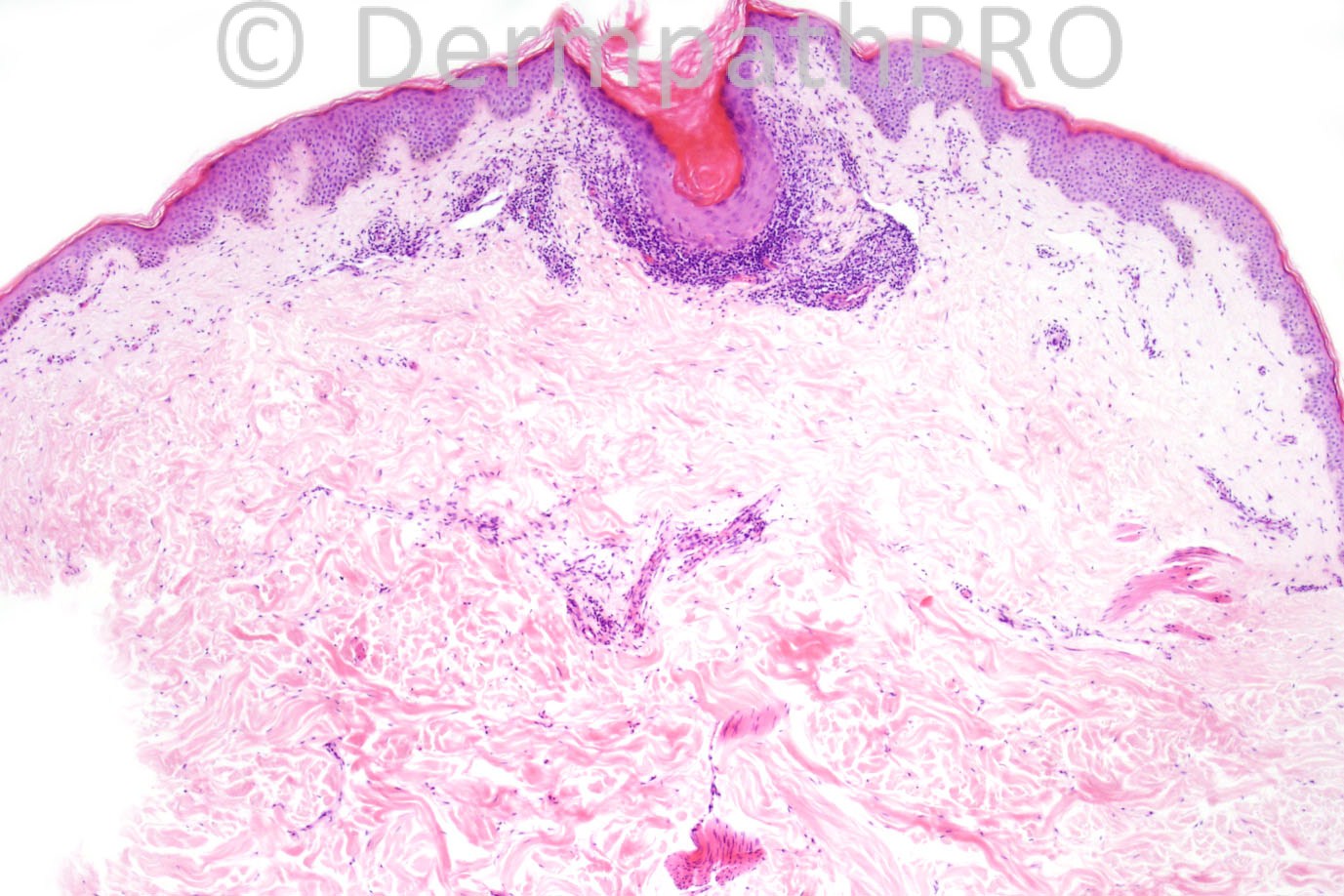
Male of 59 years with scaly small lesions on the back.


  Report Record

User Feedback


Guest Dr Engin Sezer

Posted

Follicular LP/LPP

Share this comment


Link to comment
share_externally

Dr. Mona Abdel-Halim

Posted

Follicular LP

Share this comment


Link to comment
share_externally

Robledo F. Rocha

Posted

Infundibulocentric lichenoid infiltrate of lymphocytes, infundibulum plugged by corneocytes, incipient wedge-shaped infundibular hypergranulosis and scattered necrotic keratinocytes. Interfollicular epidermis is spared. Absence of mucin between collagen bundles.
I favor lichen planopilaris.

Share this comment


Link to comment
share_externally

Guest Bansal_

Posted

lichen planopilaris

Share this comment


Link to comment
share_externally

LP/follicular LP vs lichenoid keratosis

Share this comment


Link to comment
share_externally

Guest Dr sameh abdelkodous

Posted

Lichen planopilaries

Share this comment


Link to comment
share_externally

Guest Graham Reilly

Posted

lichen planopilaris

Share this comment


Link to comment
share_externally

Dr. Phillip McKee

Posted

Lichen planopilaris is correct. The clinical history of multiple lesions on the back excludes lchenoid keratosis.

Share this comment


Link to comment
share_externally

Guest aslam parvez

Posted

Why is it not POROKERATOSIS,

Share this comment


Link to comment
share_externally

Robledo F. Rocha

Posted

[quote name='Guest' timestamp='1366049008']
Why is it not POROKERATOSIS,
[/quote]
I'm afraid to say that porokeratosis is not a good diagnostic hypothesis.
Clinically, porokeratosis tend to appear in childhood or young adulthood, and our patient here is 59 years old. Back is not a typical site for this disease, but extremities and, less frequent, sun-exposed areas. Furthermore, case history is not consistent with the characteristic gross configuration of porokeratosis, to wit, annular lesions with keratotic rim.
Microscopically, infundibular plugging is not a feature of porokeratosis, and the common denominator of all variants of this disease (cornoid lamella) is lacking.

Share this comment


Link to comment
share_externally


×
×
  • Create New...