Jump to content
In this section we have spot diagnoses posted on a daily basis since June 2010, now over 4000! You can review the archived cases and read the suggested diagnoses by users and the final comment by the contributors.
Case are uploaded each week day by 10 am UK time with the correct diagnosis will generally be posted at 8 pm UK time. Why not view the most recent spot diagnosis and proffer a diagnosis?

Case Number : Case 743 - 22 Apr Posted By: Guest

Please read the clinical history and view the images by clicking on them before you proffer your diagnosis.
Submitted Date :
   (0 reviews)

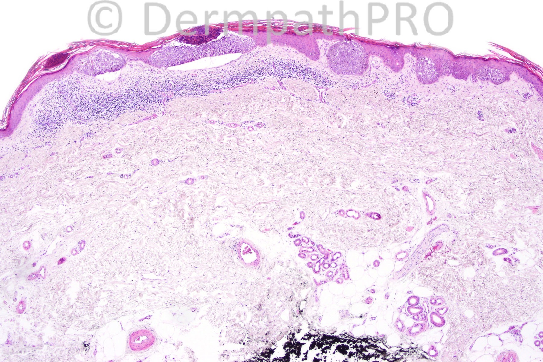
Female 56 years with a pigmented lesion on lower leg.


  Report Record

User Feedback


Multifocal superficial basal cell carcinoma

Share this comment


Link to comment
share_externally

Guest Dr Engin Sezer

Posted

Nested melanoma [i]in situ[/i]

Share this comment


Link to comment
share_externally

Guest Bansal_

Posted

On morphology, favour in situ melanoma. Would do immunohistochemistry though (S100, HMB-45, BerEP4).

Share this comment


Link to comment
share_externally

Robledo F. Rocha

Posted

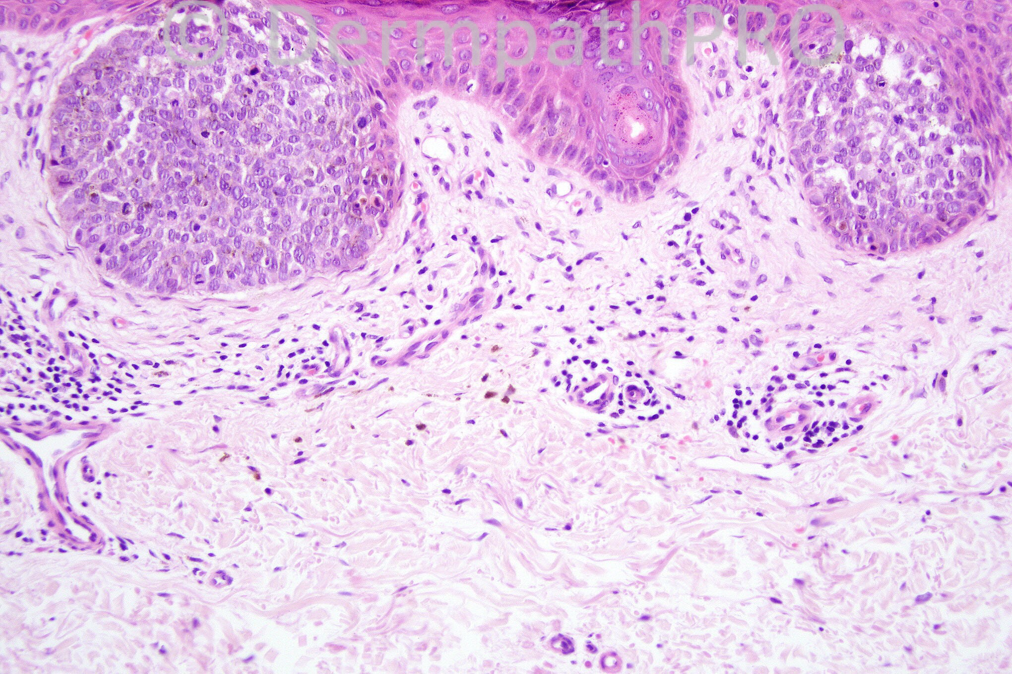
Skipping solid groups of basaloid cells attached to the epidermal undersurface showing frequent mitotic figures and peritumoral clefting. Increased mucin between tumor cells results in focal disruption. Melanin pigment can be found in basaloid cells as well as in histiocytes. Peripheral palisading is not well developed.
I think it is multifocal superficial basal cell carcinoma, pigmented type.

Share this comment


Link to comment
share_externally

Guest ANa Maria Semplici

Posted

Epitelioma basocelular superficial multifocal

Share this comment


Link to comment
share_externally

Superficial BCC, pigmented. Retraction artifact, skip lesions, stromal changes, and a hint of peripheral palisading. Not a melanocytic lesion.

Share this comment


Link to comment
share_externally

Guest Dr Engin Sezer

Posted

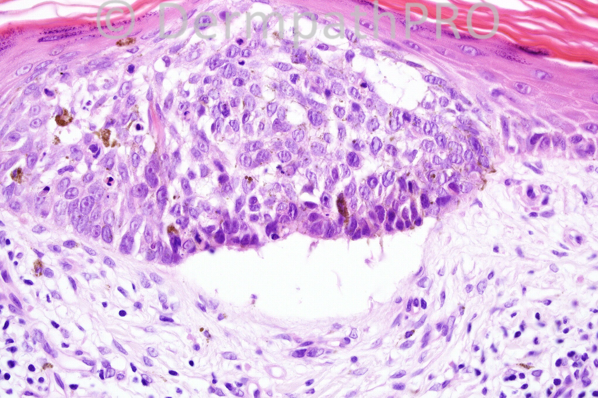
The cells in Fig.3 appear vesicular and epithelioid in appearance wirh prominent nuclei, a feature not observed in BCC.

Share this comment


Link to comment
share_externally

Dr. Richard Carr

Posted

Of course BCC can show any number of differnent phenotypes (including epithelioid cells). I favour a slightly odd melanoma like nested pattern of superficial BCC but very easily confirmed with absence of S100/melanA and positive expression of BerEP4 with completely negative EMA. The absence of pagetoid spread is very much against bowen's (clonal / pagetoid variant) and melanoma in this case. Having said that I think I have also seen exceedingly rare pagetoid cells in superficial BCC. Looking forward to seeing the answer - nice case.

Share this comment


Link to comment
share_externally

Guest Jaime Mejía MD

Posted

One way or the other I would rule out nested melanoma first by IHC.
Does the BCCS show pink prominent nucleoli? I do not know it, I favor it does not.
About the apoptosis, well, I don't know either, but theoretically it can be abolished in both cases when bcl-2 and or p53 are overexpressed.
About cleft formation, subepidermal, well, I remember some papers from Human Pathol from around 2005-6 stating the issue as a marker for melanoma, but do not know if the concept is still holding.
So, as you can tell, there's a lot of ignorance on this side, but I still think of melanoma in situ when checking the pics.

Share this comment


Link to comment
share_externally

Guest Jaime Mejía MD

Posted

Incidentally, there is some exchange of letters about the matter in the last Modern Pathology issue, referring to Dr. Kutzner description of nested melanoma in situ as an entity with epithelioid features and no pagetoid srpead. Unfortunately, access to us, USCAP members, is shutdown due to technical issues and have no more details.
Very interesting case.

Share this comment


Link to comment
share_externally

Guest Jaime Mejía MD

Posted

Kutzner et al. (2012) described a lesion that is not an in situ proper melanoma but a superficial spreading melanoma, at this moment I have to say: oops!
Looking forward to being surprised with the DX.

Share this comment


Link to comment
share_externally

Guest Dr Engin Sezer

Posted

Great comments by Dr. Mejia..

Share this comment


Link to comment
share_externally

Guest Jim Davie MD

Posted

[size=4]Pigmented superficial BCC.[/size]

[size=4]- Low-power clustered nesting pattern, lichenoid infiltrate, retraction artifact favors superficial BCC,
- A number of nests [top right photo, right-hand nest] show apparent 'eyeliner' sign with sparing of basal keratinocytes. This would weigh against in-situ MM, but raise the differential Dx of SCC in situ. There is also transepidermal nest elimination, which seems exceptional in sBCC.
- High power image shows palisading against a flat supporting BM [best seen at the left edge of the -separation cleft], that would additionally support a BCC/SCC neoplasm and disfavor Merkel or MMIS.
- Absence of significant lentiginous or intraepidermal pagetoid scatter would be rare in Bowen's or primary MMIS.[/size]

Share this comment


Link to comment
share_externally

Dr. Mona Abdel-Halim

Posted

Very nice case, very interesting discussion.
The left side nests with the minimal palisading, clefting against the basement membrane favors BCC, superficial pigmented type.
When I look to the right side nests with the lack of palisading, the presence of vesicular nuclei and prominent pink nucleoli, I feel I can not R/O large nested melanoma in situ. So, if am confronted with this, I will definitely do S100/MelanA first to exclude melanoma. Challenging...
What about a remote possibility of a collision tumor, both superficial BCC and large nested melanoma in situ !!!!!!!!!!!

Share this comment


Link to comment
share_externally

Guest Jaime Mejía MD

Posted

About collision tumor I have seen somewhere an abstract by Dr. Kutzner, I guess, about a "trichoblastomelanoma", not that this case fits the description but is an interesting possibility, I have not seen one ever in my life, but...

Share this comment


Link to comment
share_externally

Guest Rodrigo Restrepo

Posted

[color=#1C2837][font=arial, verdana, tahoma, sans-serif][size=4]Superficial basal cell carcinoma[/size][/font][/color]

Share this comment


Link to comment
share_externally

Guest Romualdo C. L. Filho

Posted

Superficial and pigmented basal cell carcinoma.

Share this comment


Link to comment
share_externally

Dr. Phillip McKee

Posted

Wow! I am thrilled by the number of comments and their excellent quality. Although this is a pigmented multifocal BCC, [i]in situ[/i] melanoma for the many reasons detailed above is an excellent differential diagnosis. However the multifocality, retraction artifact, fairly uniform basaloid cell population, absence of any interest in the overlying epidermis all favor BCC. I do not believe that any immunohistochemistry was done on this case.
[b]Next week I will be moving to live in France and will be out of contact with the web for a few days until I get my new computer. Iskander will be looking after my days for a week or so.[/b]

Share this comment


Link to comment
share_externally

Dr. Mona Abdel-Halim

Posted

Thank u so much Dr McKee, wish u a pleasant life in France

Share this comment


Link to comment
share_externally

Robledo F. Rocha

Posted

Merci pour votre constants enseignements, Dr. McKee. Je vous souhaite tout le succès possible dans votre nouvelle adresse.

Share this comment


Link to comment
share_externally

Guest Catarina Shaletich

Posted

Pigmented superficial basal cell carcinoma.

Share this comment


Link to comment
share_externally

Guest Jaime Mejía MD

Posted

Thanks Dr. McKee for sharing your expertise, experience and knowledge. I wish you the best of moving experiences, fortunately internet will allows to the privilege of contact with you from any corner in the world.

Share this comment


Link to comment
share_externally

Guest Jaime Mejía MD

Posted

[url="https://docs.google.com/file/d/0B7f20oWJtHDndFREckZxQ3ctZTA/edit?usp=sharing"]https://docs.google.com/file/d/0B7f20oWJtHDndFREckZxQ3ctZTA/edit?usp=sharing[/url]

This link above takes to a picture I have saved from the Kutzner et al paper on nested melanoma published in Mod Pathol [url="http://www.ncbi.nlm.nih.gov/pubmed/22388759#"]Mod Pathol.[/url] 2012 Jun;25(6):838-45
That's why I thought of it as a probably-possibly-maybe-not-so-sure nested melanoma, I miserably flunked :(
Dr. Kossard from Australia published a letter in Mod Pathol last ink oozing issue April 2013 stating that, well OK, there are some nested melanomas out there, he signed before as a nevoid melanoma in situ, perhaps, a novel recently identified aberrant nevi pathway. Anyway, just for the sake of not feeling depressed for the missed target.

Share this comment


Link to comment
share_externally


×
×
  • Create New...