Jump to content
In this section we have spot diagnoses posted on a daily basis since June 2010, now over 4000! You can review the archived cases and read the suggested diagnoses by users and the final comment by the contributors.
Case are uploaded each week day by 10 am UK time with the correct diagnosis will generally be posted at 8 pm UK time. Why not view the most recent spot diagnosis and proffer a diagnosis?

Case Number : Case 744 - 23 Apr Posted By: Guest

Please read the clinical history and view the images by clicking on them before you proffer your diagnosis.
Submitted Date :
   (0 reviews)

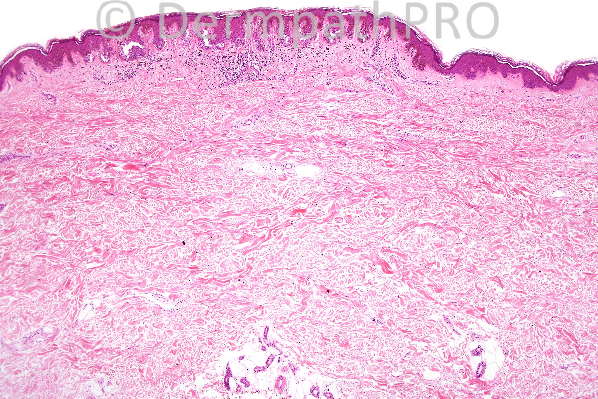
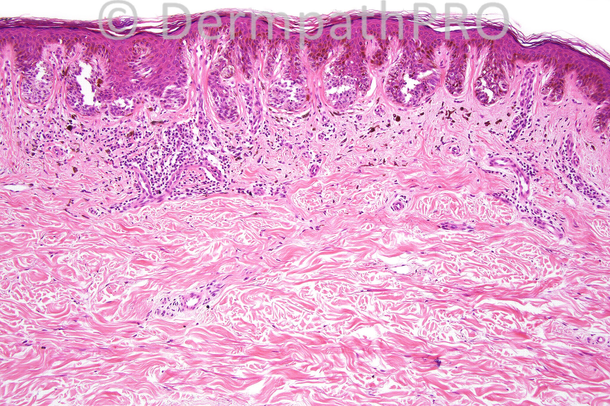
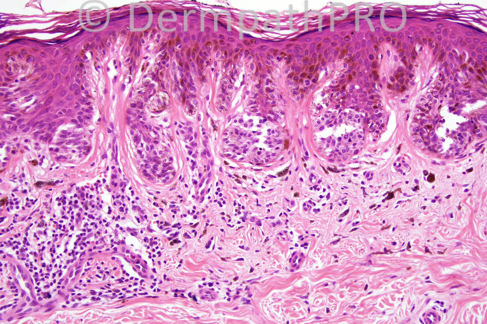
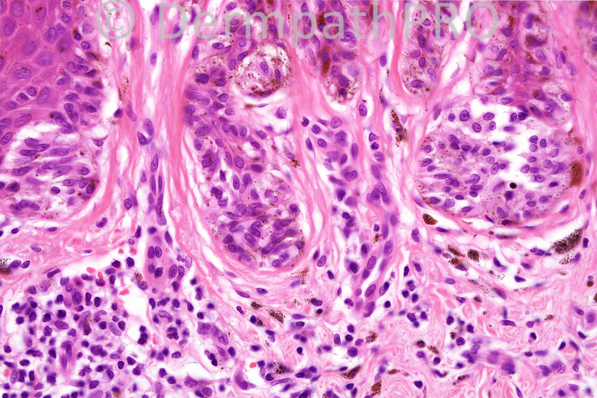
Male 39 years with a pigmented lesion in the popliteal fossa.


  Report Record

User Feedback


Guest Bansal_

Posted

Melanocytic lesion with architectural (lamellar fibroplasia, bridging) and minimal / mild cytological atypia. No pagetoid spread and I cannot identify mitoses. Would call this a mildly dysplastic naevus.

Share this comment


Link to comment
share_externally

Guest Romualdo C. L. Filho

Posted

I can't see random cytologic atypia of any significant degree. I would call this lesion lentiginous junctional melanocytic nevus.

Share this comment


Link to comment
share_externally

Guest Rodrigo Restrepo

Posted

Mild dysplastic naevus

Share this comment


Link to comment
share_externally

Guest Dr. Francisco Vílchez

Posted

Nevus

Share this comment


Link to comment
share_externally

Robledo F. Rocha

Posted

Nested and lentiginous atypical melanocytic proliferation. Nests vary in size and shape, and some tend to be irregularly located at the side of rete ridges. Some nests are horizontally oriented with focal bridging (Picture #2, left). Host response takes form of lymphocytic infiltrate and lamellar fibroplasia. Cytologic atypia is mild, that melanocytic nuclei are as large as keratinocytes nuclei.
There’s no intradermal component.
I think it's a low-grade junctional dysplastic nevi.

Share this comment


Link to comment
share_externally

Lentiginous junctional dysplastic nevus, mild. Slight architectural disorder with nests at tips or rete and creeping up the sides, peri-rete lamellar fibrosis, minimal cytologic atypia, and a mild host immune response - all features of a mildly dysplastic nevus.

Share this comment


Link to comment
share_externally

Guest Jim Davie MD

Posted

[font=georgia,serif][size=3]Ah, nothing so factious as interpretation of the humble benign nevus. Don't think there will be any wrong answers on this one! ;)

I favor a mildly dysplastic junctional nevus, likely arising in preexisting nevus.
The differential dx would be a 'nevus of special site' (flexural area) given popliteal location. The inflammation is the main differentiator for me to push it to dysplastic (likely arising in pre-existing special-site junctional nevus, given the far lateral nests that appear uninvolved).

[b]Benign nevus of special site features:[/b]
- Nests are slightly irregular in size and placement, but predominantly at tips of rete pegs.
- No significant rete bridging (there seem to be keratinocytes involved at all junctures, therefore I think these are tangential 'pseudobridging').
- Overall bilateral symmetry. Tapering edges, and widely scattered edge nests. Lentiginous component is centrally restricted.[/size][/font]
[b]Dysplastic features:[/b]
[font=georgia,serif][size=3]- Slight periretal fibrosis
- Targeted lichenoid inflammation.
- Melanocytes withlow-grade atypia (at best): roughly the same size as basal keratinocytes in nuclear size, even chromatin, lack of hyperchromatism, pink cytoplasm with coarse/giant melanosomes, focally prominent nucleoli, lack of proliferative activity.[/size][/font]

Share this comment


Link to comment
share_externally

Guest Giedrius Salkus

Posted

This lesion occurs on a flexural site and that makes differential diagnosis tricky. However in my opinion, this melanocytic lesion meets the criteria for dysplastic nevus, as there is architectural atypia, dermal response and one can always find slight cytological atypia when needed. It is completely removed, and that is ok. I would hesitate in case margin was involved, but would probably advice narrow reexcision. This is a low grade lesion, but not as low I could sleep well.

Share this comment


Link to comment
share_externally

junctional dysplastic nevus vs site specific

Share this comment


Link to comment
share_externally

I see nested and few lentiginous melanocytic proliferation with some atypia. The nests are located at the side and tip of rete ridges. I did not see bridging. I think it is a special site benign melanocytic nevus.

Share this comment


Link to comment
share_externally

Dr. Mona Abdel-Halim

Posted

I will call it mildly dysplastic junctional nevus.

Share this comment


Link to comment
share_externally

Guest Jaime Mejía MD

Posted

Junctional melanocytic nevus from special-site type (flexural site)[list]
[*]architectural atypia
[*]no cytological dyplasia
[*]discohesiveness
[/list]

Share this comment


Link to comment
share_externally

Guest Dr. Marcela M. Escanamé

Posted

atypical junctional nevus

Share this comment


Link to comment
share_externally

Dr. Phillip McKee

Posted

Again, it is great to see so many comments. I think that this is a difficult lesion to interpret and the differential diagnosis certainly lies between a flexural-type nevus and a dysplastic nevus. When I photographed the case I particularly included figure 4 to show at high power that the lesion was very much a nested lesion with the nests residing at the tips of the rete ridges. There is very little in the way of a lentiginous element. The architectural disorder of a dysplastic nevus is therefore absent. I do not think that there is any real cytological atypia although its presence would not really distinguish between the two entities. The stromal changes are common to both. My diagnosis therefore rests with the growth pattern and stromal changes and I regard this lesion as a nevus of special sites (flexural type). I must comment that such nevi may be seen just about anywhere and therefore the term should one day become redundant. I would not be concerned if people called this a junctional nevus! Jim Davie sumarized the situation quite nicely with his first sentence. I think that it is important not to call this lesion a dysplastic nevus because of the implications for the patient (unless of course you don't believe in the concept of dysplastic nevus!!!).

PS I will be back on Mondays and Tuesdays in a couple of weeks.

Share this comment


Link to comment
share_externally

Robledo F. Rocha

Posted

[quote name='Jim Davie MD' timestamp='1366724177']
[font=georgia, serif][size=3]Ah, nothing so factious as interpretation of the humble benign nevus. Don't think there will be any wrong answers on this one! ;)[/size][/font]
[/quote]
Just perfect, Dr. Davie!

Share this comment


Link to comment
share_externally


×
×
  • Create New...