Jump to content
In this section we have spot diagnoses posted on a daily basis since June 2010, now over 4000! You can review the archived cases and read the suggested diagnoses by users and the final comment by the contributors.
Case are uploaded each week day by 10 am UK time with the correct diagnosis will generally be posted at 8 pm UK time. Why not view the most recent spot diagnosis and proffer a diagnosis?

Case Number : Case 747 - 26 Apr Posted By: Guest

Please read the clinical history and view the images by clicking on them before you proffer your diagnosis.
Submitted Date :
   (0 reviews)

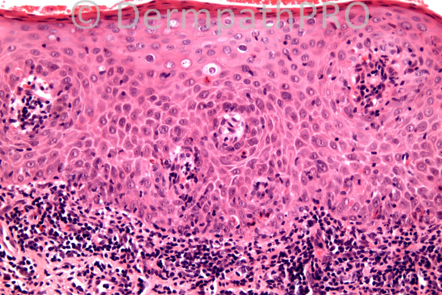
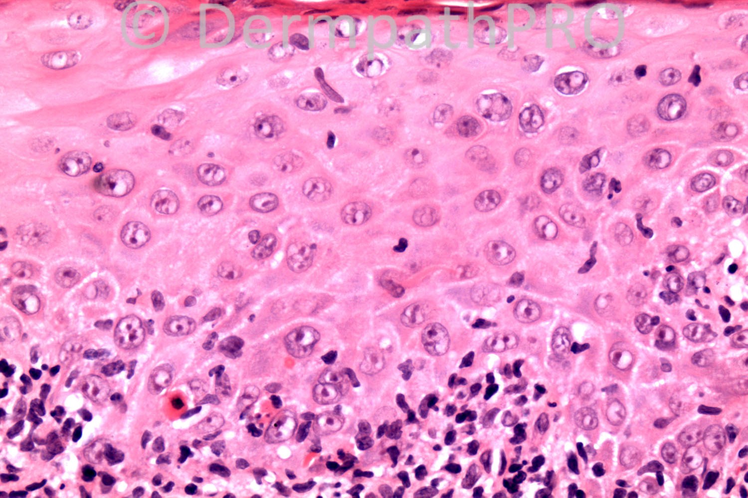
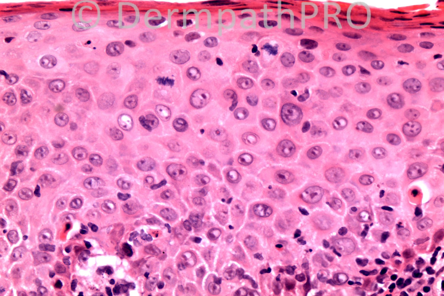
49 years-old male. Inflammatory macule ?eczema ?psoriasis.

Case posted by Dr. Richard Carr.

  Report Record

User Feedback


Dr. Mona Abdel-Halim

Posted

Bowen's disease.

Share this comment


Link to comment
share_externally

Dr. Richard Carr

Posted

Thanks Mona. The anatomical site is glans penis I think.

Share this comment


Link to comment
share_externally

Dr. Mona Abdel-Halim

Posted

If so, then it will be erythroplasia of Queyrat !!!!!

Share this comment


Link to comment
share_externally

Guest Bansal_

Posted

Dysplasia / PeIN but unsure of grading. In real-life, would take more levels and show to colleagues.

Share this comment


Link to comment
share_externally

Robledo F. Rocha

Posted

Differentiated penile intraepithelial neoplasia (PeIN), probably arising in the setting of a long-term dermatitis.
I prefer the simplified nomenclature for penile preinvasive lesions proposed by Antonio Cubilla et al (Morphological characterization and distribution of penile precancerous lesions using a simplified nomenclature. A study of 198 lesions in 115 patients. Lab Invest 2008;88:696A).

Share this comment


Link to comment
share_externally

Guest Rodrigo restrepo

Posted

[color=#1C2837][font=arial, verdana, tahoma, sans-serif][size=4]Differentiated PeIN[/size][/font][/color]

Share this comment


Link to comment
share_externally

Guest Jim Davie MD

Posted

Agree with SCC in situ / Bowen's disease / Erythoplasia of Queyrat (The tree of nomenclature needs pruning!)
[color=#000080] - [i]Low power image (top left)[/i]: shows increased pleomorphism and nuclear/cytoplasm volumes, compared to the 'normal' epidermis on the left, and what look like sparse bizarre giant keratinocytes and deeply eosinophilic necrotic keratinocytes in the upper reaches. [very useful image].[/color]
[color=#000080] - [i]Medium and high power images (top right, lower right)[/i]: show mitotic figures at all levels, coarse parakeratosis, dyskeratosis, and prominent nucleoli. There is aberrant multinucleation.[/color]

Share this comment


Link to comment
share_externally

Dr. Richard Carr

Posted

[size=3][font=Arial]Thank you all for the contributions. I must say that the contributors today all made light work of a difficult case which is very impressive. I am saying this so that any visitors new to dermatopathology (the neophytes) will not be disheartened!! Immunostaining can be exceedingly useful in this setting. [/font][/size]

[size=3][font=Arial]In view of the comments above I e-mailed to Dr Cubilla as follows:[/font][/size]

[size=3][font=Arial]Dear Dr Cubilla,[/font]
I would be most interested on your opinion on the case I posted today for Phillip Mckee's website - the spot diagnosis of the day. I reported it as [b]conventional [high grade] PeIN [/b]because of strong p16 and what I interpreted as focal koilocytosis. I thought the lichenoid reaction was limited to the neoplastic epithelium and was therefore secondary but I would be most grateful for your thoughts.
Richard Carr

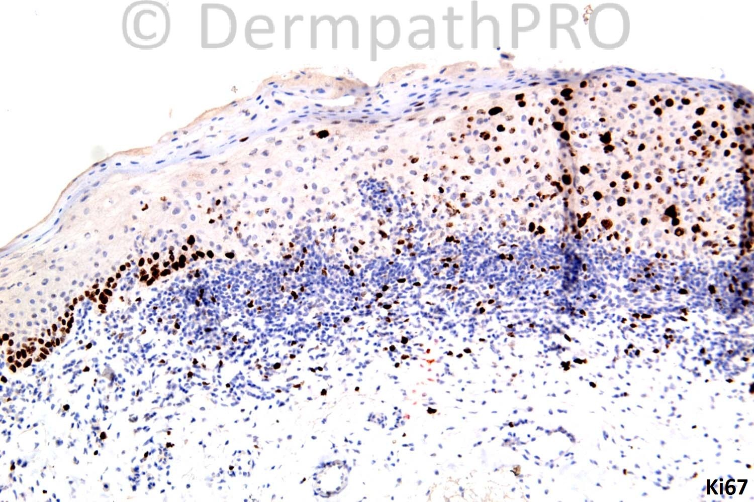
Dr Cubilla responds: I would favor this lesion to be a Penile Intraepithelial Neoplasia,basaloid over Diff. PeIN. Cells are rather uniform, surface is parakeratotic with isolated superficial koylocytotic cells. I expect this lesion to be p16 Ki 67 positive. Thanks for calling my attention to this intersting case. Antonio Cubilla.

Additional notes (Dr Carr): Abrupt full thickness p16 with Ki67 at all levels coincides with the inflammatory infiltrate (on the right half of image 1). [/size]

[size=3]I will ask the DermpathPro team to post the additional immunos for your interest. Regards to all and enjoy your weekends[/size]

Share this comment


Link to comment
share_externally

Guest Bansal_

Posted

Thanks Richard, very useful.

Share this comment


Link to comment
share_externally

Dr. Mona Abdel-Halim

Posted

Thanks for the Immunos , very nice

Share this comment


Link to comment
share_externally


×
×
  • Create New...