Jump to content
In this section we have spot diagnoses posted on a daily basis since June 2010, now over 4000! You can review the archived cases and read the suggested diagnoses by users and the final comment by the contributors.
Case are uploaded each week day by 10 am UK time with the correct diagnosis will generally be posted at 8 pm UK time. Why not view the most recent spot diagnosis and proffer a diagnosis?

Case Number : Case 748 - 29 Apr Posted By: Guest

Please read the clinical history and view the images by clicking on them before you proffer your diagnosis.
Submitted Date :
   (0 reviews)

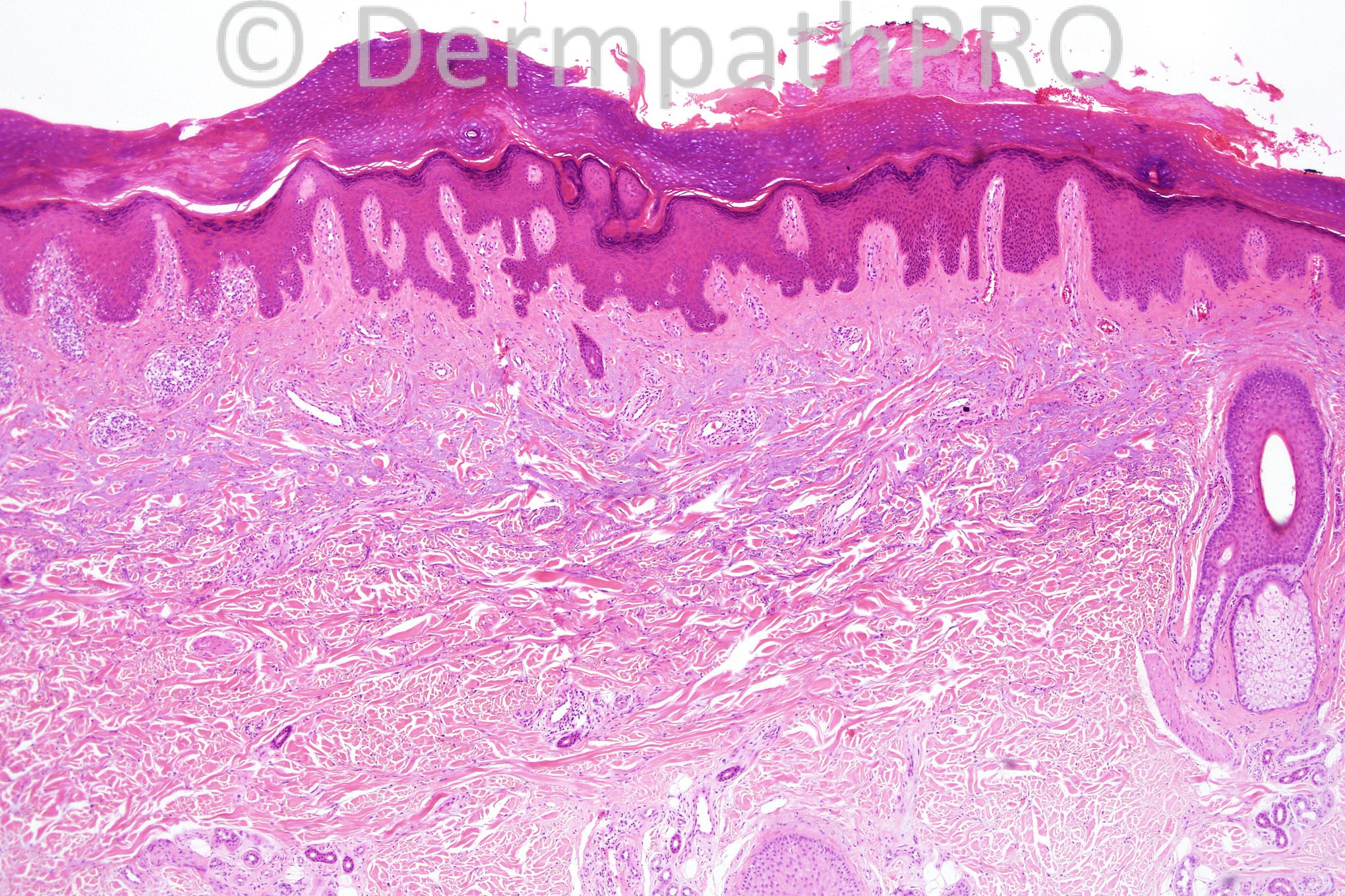
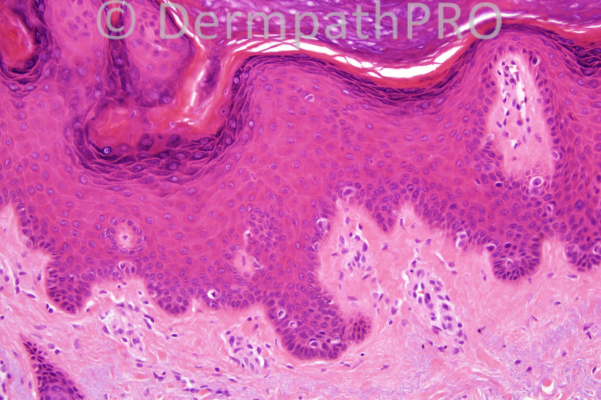
Female 52 years with a scaly plaque on her arm.


  Report Record

User Feedback


Guest Dr Engin Sezer

Posted

Lichen simplex

Share this comment


Link to comment
share_externally

Guest nick turnbull

Posted

pagetoid reticulosis?

Share this comment


Link to comment
share_externally

Dr. Mona Abdel-Halim

Posted

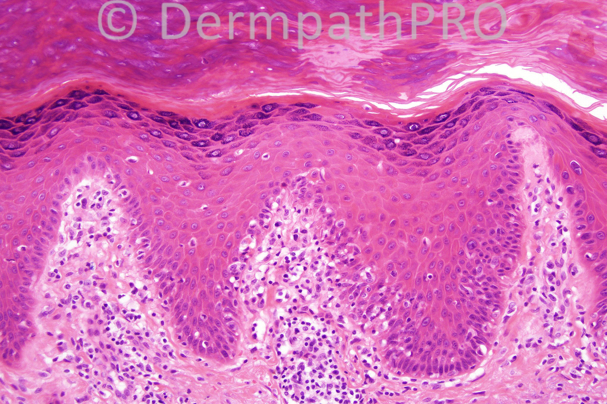
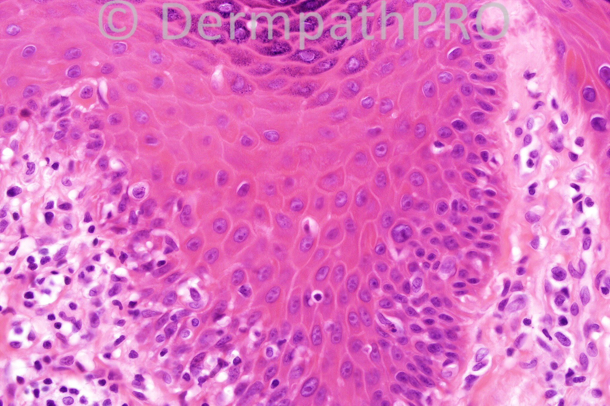
I can see marked orthokeratosis with focal parakeratosis on the right side. The 3rd and 4th images illustrates with certainty an interface reaction with necrotic keratinocytes along the basal cell layer and above it. There is marked solar elastosis. The lesion is single as appears from the clinical description. I am thinking of the rare creeping form of LPLK, which shows only focal acute activity. Although lichen simplex may explain the apparently thickened collagen in the dermal papillae and the marked orthokeratosis but it does not explain the interface dermatitis seen focally. I think itching can cause some scattered necrotic keratinocytes but not as this peculiar pattern seen here along the DEJ associated with interface lymphocytes. Nice case...

Share this comment


Link to comment
share_externally

Dr. Richard Carr

Posted

Please take a look at the extra immuno images on last Friday's case (747) that the Dermpath team posted for me. Shows how helpful immuno can be at times!

Share this comment


Link to comment
share_externally

Guest Jim Davie MD

Posted

[size=4][font=arial, helvetica, sans-serif]Looks to me like three processes:
1.[i] Lichen simplex chronicus[/i]: [color=#000080]Primary findings are psoriasiform hyperplasia with hyperorthokeratosis, wedge-shaped hypergranulosis, stratum lucidum, and mixed acral type basophilic compact hyperorthokeratosis and parakeratosis, with mild acanthosis of a terminal hair infundibulum. Rare necrotic keratinocytes and sparse spongiosis are expected.[/color]
2. [i]Chronic inflammatory reaction[/i]: [color=#000080] Mixture of solitary, small lymphocyte-like hyperchromatic cells and larger mononuclear cells with nucleoli and open nuclei, some with perinuclear halos, with patchy asymmetric distribution in epidermis and papillary dermis. There is predominantly low level pagetoid scatter, with occasional elimination into the stratum corneum. This looks like a classic inflammatory reaction pattern in LSC, to me. Pagetoid reticulosis would be a DDx consideration, (as it often presents with psoriasiform architecture), but the mixed cytology and low density patchy infiltrate doesn't make it for me. Stains for CD4 and CD8 (should be CD4+ in most cases), and CD1a for Langerhans' cells might clear up the identity of the intraepidermal infiltrate if there is doubt. [/color]
3. [i]Marked solar elastosis[/i].[/font][/size]

Share this comment


Link to comment
share_externally

I suspect for LSC. In Pagetoid reticulosos (PG) i wolud expect more epidermotropism. I also think we would expect lots of CD30 cells.

Share this comment


Link to comment
share_externally

Iskander H. Chaudhry

Posted

Thanks for everyone who contributed, the diagnosis is lichen simplex chronicus.

Share this comment


Link to comment
share_externally


×
×
  • Create New...