Jump to content
In this section we have spot diagnoses posted on a daily basis since June 2010, now over 4000! You can review the archived cases and read the suggested diagnoses by users and the final comment by the contributors.
Case are uploaded each week day by 10 am UK time with the correct diagnosis will generally be posted at 8 pm UK time. Why not view the most recent spot diagnosis and proffer a diagnosis?

Case Number : Case 901 - 2nd December Posted By: Guest

Please read the clinical history and view the images by clicking on them before you proffer your diagnosis.
Submitted Date :
   (0 reviews)

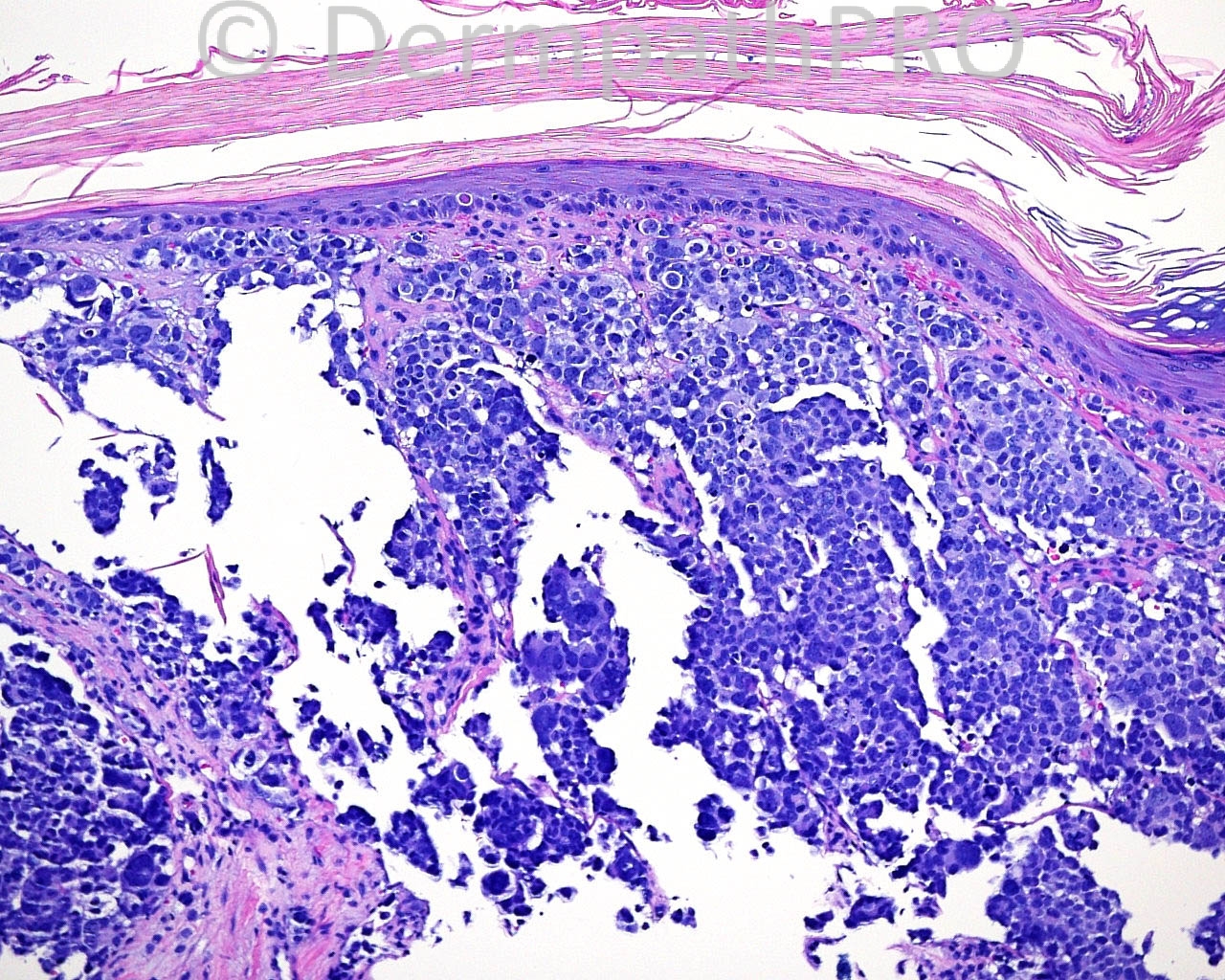
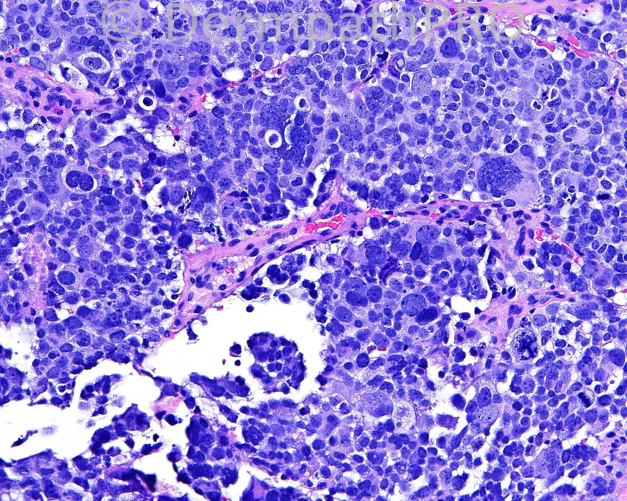
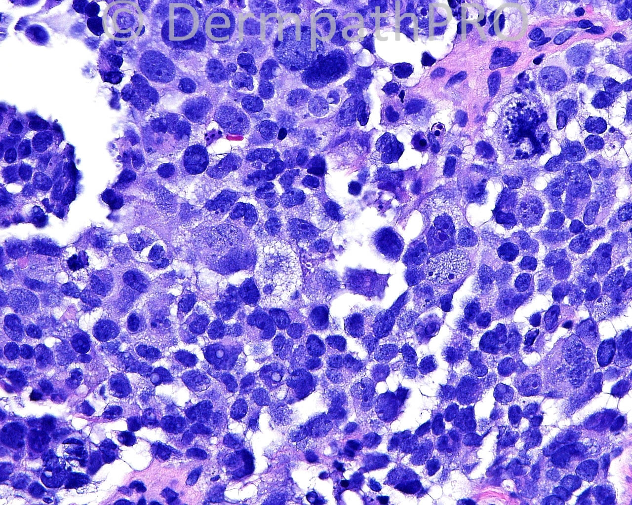
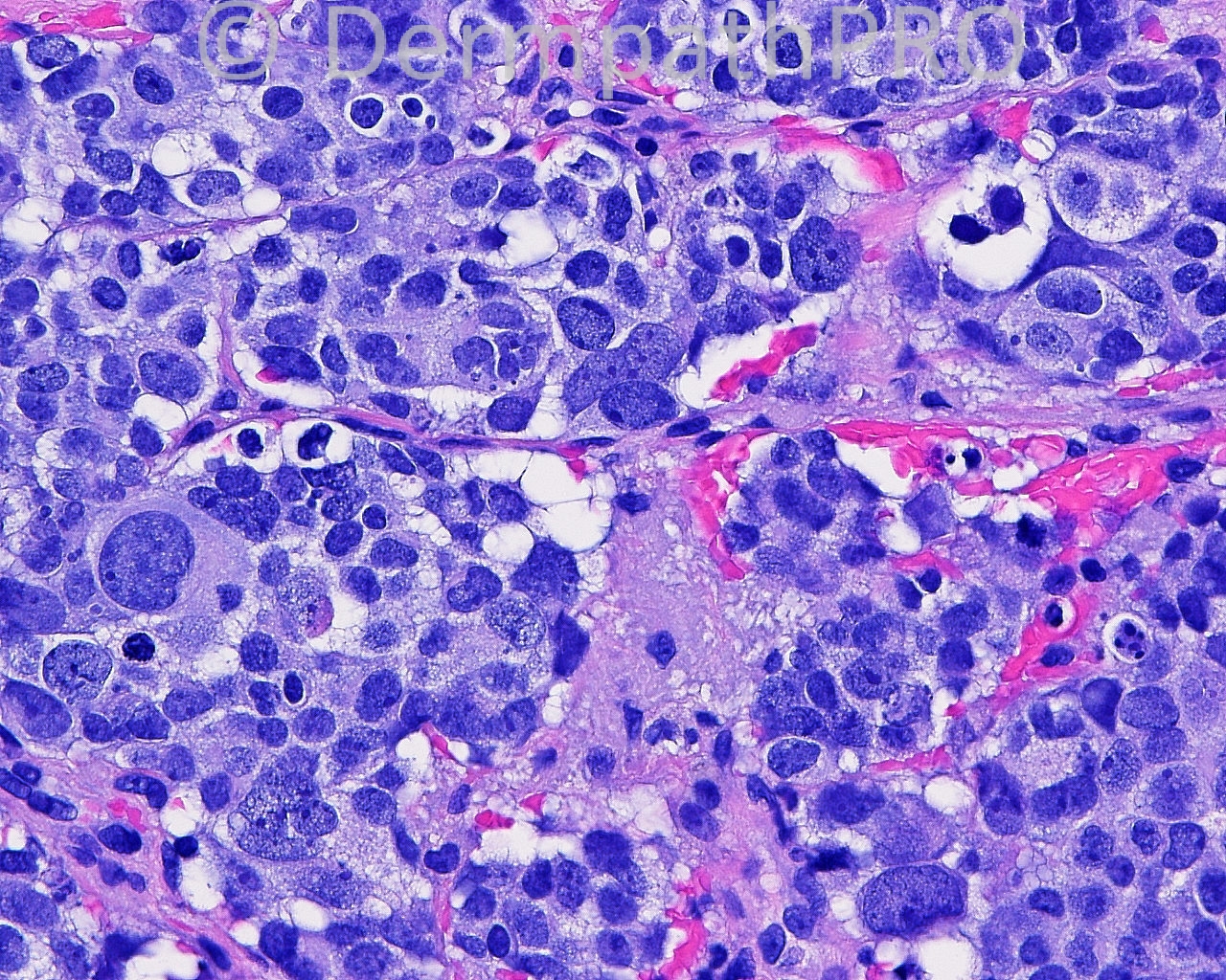
The patient is an 85 year old white man with a history of a lesion that was excised from the nose, with known outcome. He was treated. A shave biopsy of a keratotic, pink papule arising in the flat used to close the nose defect, present for three months, is taken from the nose.

Case posted by Dr. Mark Hurt


  Report Record

User Feedback


Guest Maria George

Posted

Recurrent malignant melanoma is my favorite here however others need to be excluded by IHC.

Share this comment


Link to comment
share_externally

Dr. Richard Carr

Posted

Friday's case was a metastasis from the colon (CDX2, CEA positive). Apologies for delay, long day in Paris!

Share this comment


Link to comment
share_externally

I also think it is a recurrent Melanoma. If there is no history, immunohistochemistry is mandatory.

Share this comment


Link to comment
share_externally

Guest Juan Carlos Garcés, Ecuador

Posted

Agree with recurrent melanoma

Share this comment


Link to comment
share_externally

Robledo F. Rocha

Posted

Pleomorphic recurrence of melanoma or squamous cell carcinoma.

Share this comment


Link to comment
share_externally

Guest Jim Davie MD

Posted

I favor a high-grade squamous carcinoma. There is high-grade atypia at the junction, most pronounced / best seen in the first low-power image (right edge). Given the epidermal atypia, and clinical history, there is consideration that this tumor is an 'import' from the flap, and not original to the nose.

To add to the above differential of SCC vs. melanoma, less likely would be:
1. Epithelioid angiosarcoma ( given other images, it seems that the red cells in last photo are most likely artefactual to procedure, but some appear placed within intracellular lumens. )
2. High grade sebaceous carcinoma (the vacuolar clear-cell changes seem more consistent with that commonly seen in poorly differentiated SCC / Bowen's disease).

Share this comment


Link to comment
share_externally

Dr. Mona Abdel-Halim

Posted

I also thought of recurrent sebaceous carcinoma, but the vague nested nature of the tumor masses made me favor melanoma. I have realized the atypia of the basal layer but explained it by actinic keratosis in the flap, the tumor masses did not look like squamous cell carcinoma to me, but it is of course a possibility... I still feel it is a recurrence of the original tumor.. Lovely case...

Share this comment


Link to comment
share_externally

Dr. Richard Carr

Posted

Don't forget metastasis (from a systemic primary) is also a possibility. What looks like nesting pattern may be intra-vascular tumour. Certainly some endothelial looking cells noted around some nests. Agree should consider all of the above (including angiosarcoma).

Share this comment


Link to comment
share_externally

Eman El-Nabarawy

Posted

Favor recurrent sebaceous carcinoma with a DD of melanoma.

Share this comment


Link to comment
share_externally

Eman El-Nabarawy

Posted

Merkel cell carcinoma (neuroendocrine carcinoma).

Share this comment


Link to comment
share_externally


Join the conversation

You can post now and register later. If you have an account, sign in now to post with your account.

Guest
Add a comment...

×   Pasted as rich text.   Paste as plain text instead

  Only 75 emoji are allowed.

×   Your link has been automatically embedded.   Display as a link instead

×   Your previous content has been restored.   Clear editor

×   You cannot paste images directly. Upload or insert images from URL.

Loading...

×
×
  • Create New...