Jump to content
In this section we have spot diagnoses posted on a daily basis since June 2010, now over 4000! You can review the archived cases and read the suggested diagnoses by users and the final comment by the contributors.
Case are uploaded each week day by 10 am UK time with the correct diagnosis will generally be posted at 8 pm UK time. Why not view the most recent spot diagnosis and proffer a diagnosis?

Case Number : Case 903 - 4th December Posted By: Guest

Please read the clinical history and view the images by clicking on them before you proffer your diagnosis.
Submitted Date :
   (0 reviews)

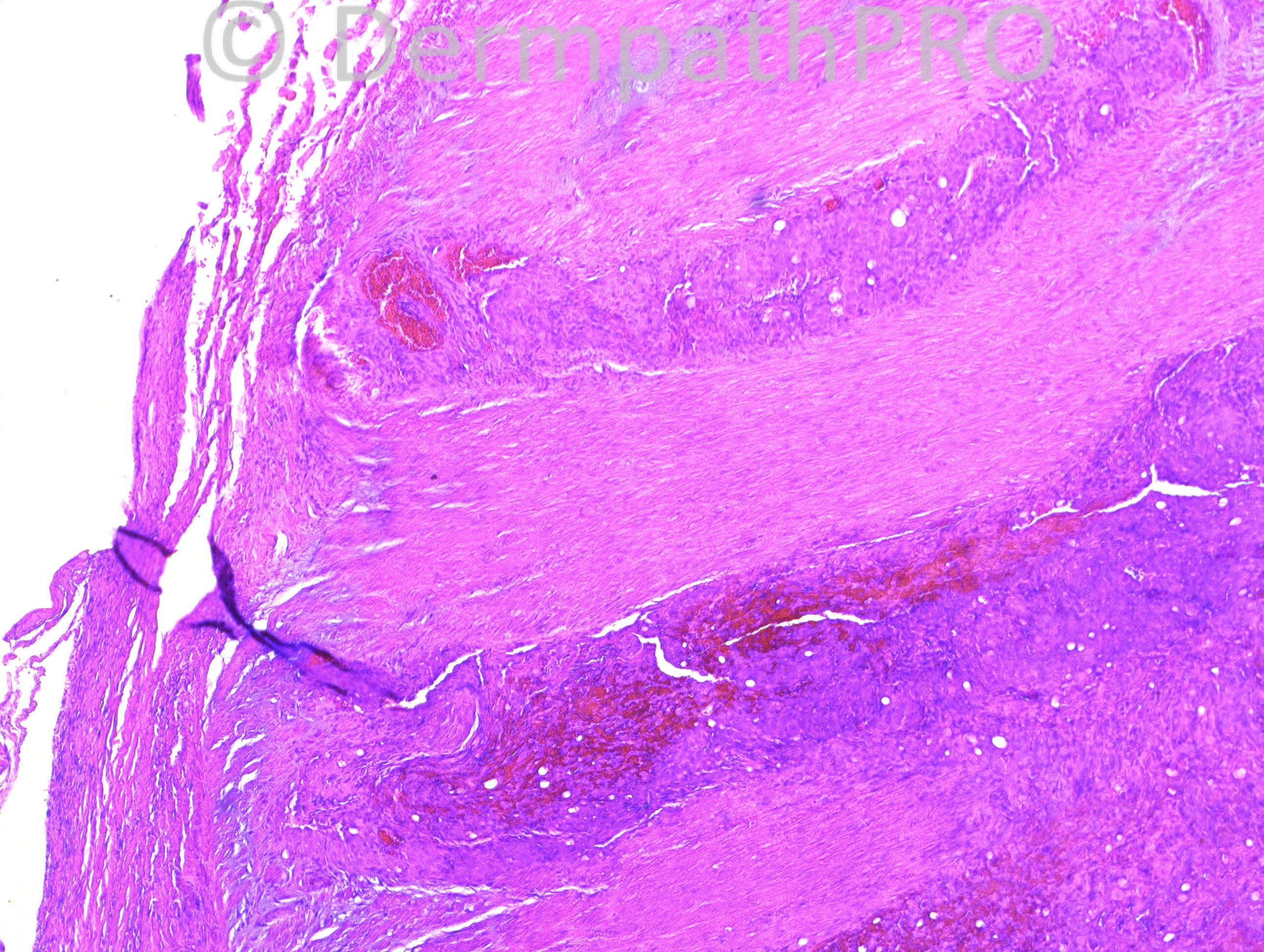
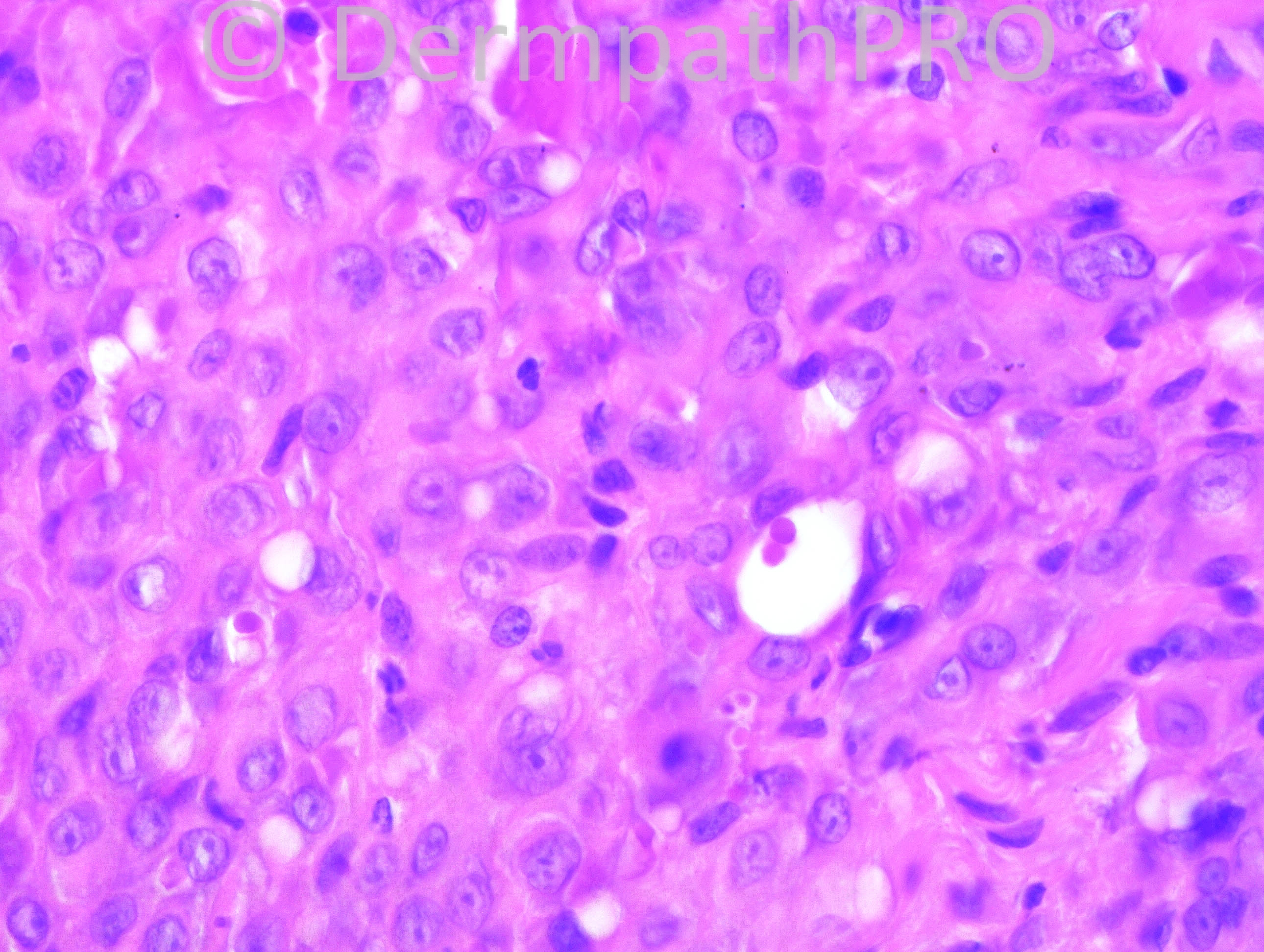
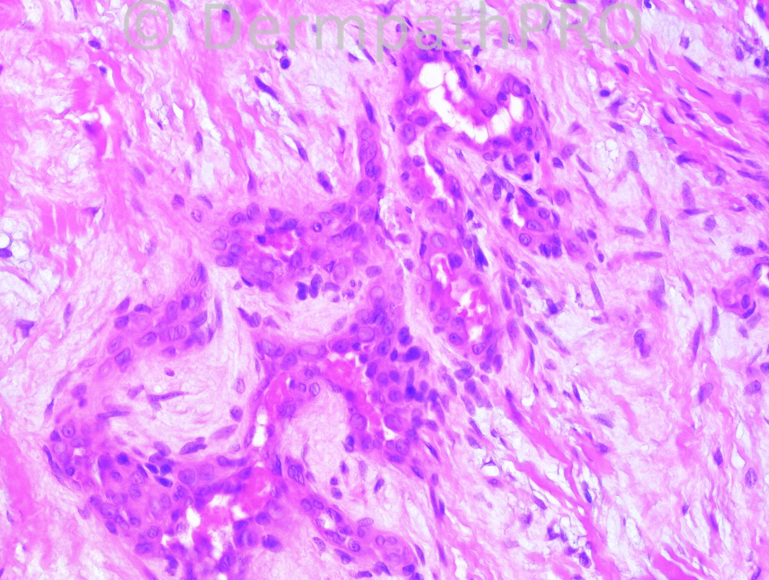
48-year-old female with left wrist mass.

Case posted by Dr. Hafeez Diwan.


  Report Record

User Feedback


Hanan Vaknine MD

Posted

[font="StoneSerif"][size="1"][font="StoneSerif"][size="1"]Epithelioid hemangioendothelioma[/size][/font][/size][/font]

Share this comment


Link to comment
share_externally

Guest Mazen charaf iddin

Posted

agree [font=StoneSerif][size=1][size=1]Epithelioid hemangioendothelioma[/size][/size][/font]

Share this comment


Link to comment
share_externally

Epithelioid hemangioendothelioma. Don´t see malignant features here.

Share this comment


Link to comment
share_externally

Guest Juan Carlos Garcés, Ecuador

Posted

Epithelioid hemangioendothelioma

Share this comment


Link to comment
share_externally

Guest Maria George

Posted

Epitheloid Sarcoma.
suggested IHC panel ;CK, CD34, iNI1

Share this comment


Link to comment
share_externally

Guest Romualdo

Posted

Except for the lower power view all images show us an epithelioid vascular neoplasia. There are not obvious signs of malignancy so I think epithelioid angiosarcoma can be excluded. Although I cannot see eosinophils there are some lymphocytes in the last figure and the well formed vessels seen in figure 4 are not a characteristic of epithelioid hemangioendotheliomas. So, I think this is an epithelioid hemangioma.

Share this comment


Link to comment
share_externally

Robledo F. Rocha

Posted

Intratumoral extravasated erythrocytes make me think of vascular tumor. In the face of lacking brisk mitotic activity within solid sheets of epithelioid cells with intracytoplasmic lumens, I go with epithelioid hemangioendothelioma.

Share this comment


Link to comment
share_externally

Dr. Mona Abdel-Halim

Posted

The vascular spaces in the 4th image as Romualdo said are not seen in EHE. I am also thinking of epithelioid hemangioma, may be esinophils are present elsewhere!!!!! I am a bit puzzled with the first image, could it be an intravascular epithelioid hemangioma???

Share this comment


Link to comment
share_externally

Guest Jim Davie MD

Posted

[size=4][font=arial,helvetica,sans-serif]I agree with epithelioid hemangioendothelioma vs. hemangioma. The atypia level and mitotic level is low, so doubt angiosarcoma or epithelioid sarcoma.

[i]Other differential diagnosis considerations:[/i][/font][/size]
[font=arial,helvetica,sans-serif][size=4]1. Retiform hemangioendothelioma has curvilinear arcuate vessels with hobnailing, and collagenous fibrosis, but not the solid areas, seen in these images. The clinical presentation (extremity of adult) would be supportive.
2. Masson's tumor (intravascular epithelial hyperplasia), would be in the differential for a low-grade vascular proliferation lacking significant mitotic activity. Clinical history (hand lesion, middle-age female) would be a very common presentation.
3. Kaposi's sarcoma. [/size][/font]

Share this comment


Link to comment
share_externally

Guest Rodrigo Restrepo

Posted

[color=#1C2837][font=arial, verdana, tahoma, sans-serif][size=4]Epithelioid hemangioendothelioma[/size][/font][/color]

Share this comment


Link to comment
share_externally

Guest Maria George

Posted

Epitheid sarcoma-lke hemangioendthelioma is agood posibiliy here.

Share this comment


Link to comment
share_externally

Dr. Hafeez Diwan

Posted

I called this epithelioid hemangioendothelioma.

Share this comment


Link to comment
share_externally


Join the conversation

You can post now and register later. If you have an account, sign in now to post with your account.

Guest
Add a comment...

×   Pasted as rich text.   Paste as plain text instead

  Only 75 emoji are allowed.

×   Your link has been automatically embedded.   Display as a link instead

×   Your previous content has been restored.   Clear editor

×   You cannot paste images directly. Upload or insert images from URL.

Loading...

×
×
  • Create New...