Jump to content
In this section we have spot diagnoses posted on a daily basis since June 2010, now over 4000! You can review the archived cases and read the suggested diagnoses by users and the final comment by the contributors.
Case are uploaded each week day by 10 am UK time with the correct diagnosis will generally be posted at 8 pm UK time. Why not view the most recent spot diagnosis and proffer a diagnosis?

Case Number : Case 906 - 9th December Posted By: Guest

Please read the clinical history and view the images by clicking on them before you proffer your diagnosis.
Submitted Date :
   (0 reviews)

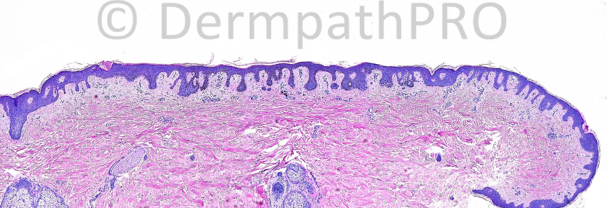
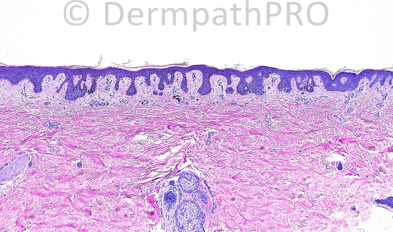
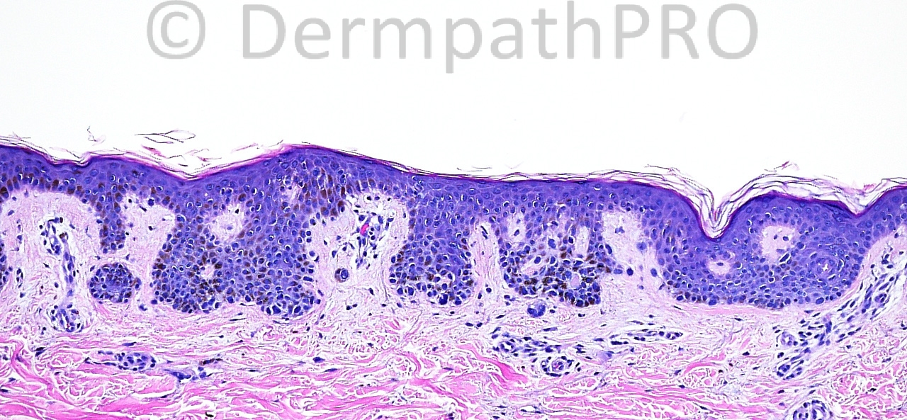
The patient is a 34 year old woman with an excision of a lesion on the right upper back.

Case posted by Dr. Mark Hurt


  Report Record

User Feedback


Sasi Attili

Posted

Presuming this is the worse it gets on all levels through the specimen- I think, this is something I would do S100/ Melan A stains to see if there is a true melanocytic proliferation or if the pigmentation is simply within the keratinocytes. Early flat/ reticulated pigmented BCP can often overlap (or be difficult to distinguish) from a solar lentigo. Histo is reminiscent of that.

Share this comment


Link to comment
share_externally

My first thought was a Becker nevus.

I can see some epidermal hyperplasia with anastomosing rete ridges and basal hyperpigmentation.

Share this comment


Link to comment
share_externally

Robledo F. Rocha

Posted

Hyperpigmented rete ridges that are too elongated and anastomosing, accompanied by some basophilic elastic fibers, make me think of solar lentigo evolving into reticulated seborrheic keratosis. Patient and clinical dermatologist, maybe concerned with the grossly modifications brought by this process, have decided to remove the lesion.
I didn’t see significant increased numbers of melanocytes, so that pigmented cell are keratinocytes to me.

Share this comment


Link to comment
share_externally

Dr. Mona Abdel-Halim

Posted

Solar lentigo starting to evolve into retic seb K, do not c nests to think of a junctional nevus..

Share this comment


Link to comment
share_externally

Dr. Mona Abdel-Halim

Posted

I think the second Melan A is highlighting nests that I did not perceive on H&E, this will make it a lentiginous junctional nevus.

Share this comment


Link to comment
share_externally

Sasi Attili

Posted

Wow. Impressive lentiginous melanocytic proliferation. It does look benign, but I am not sure it fits into the category of 'junctional nevus'. I am not convinced that the nest in the dermis is indeed dermal and not just a cross cut rete ridge. I don't see any pagetoid spread at all to call this lesion definitely malignant, though the melanocytic proliferation is quite confluent. Tough call. I would however favour this being within the Benign Junctional Nevus spectrum.

Share this comment


Link to comment
share_externally

Dr. Richard Carr

Posted

Junctional / lentiginous naevus ("jentigo")

Share this comment


Link to comment
share_externally

Guest Jim Davie MD

Posted

Lentiginous compound nevus. I think at least some of the sparse superficial Melan-A positive cells near the junction may be actual dermal melanocytes rather than tangential sections of rete. The junctional melanocytes cluster along tips of rete pegs, and predominantly spares the intra-rete zones. No high-grade atypia...melanocytes are predominantly small with rounded/hyperchromatic nuclei. ( There is background of subtly lighter-brown, non-staining heavily pigmented keratinocytes and focally aggregated melanophages ).

Clinically, I think it would have been any darkly pigmented lesion, such as dark area in a nevus spilus (r/o MM), or inkspot lentigo.

Share this comment


Link to comment
share_externally


Join the conversation

You can post now and register later. If you have an account, sign in now to post with your account.

Guest
Add a comment...

×   Pasted as rich text.   Paste as plain text instead

  Only 75 emoji are allowed.

×   Your link has been automatically embedded.   Display as a link instead

×   Your previous content has been restored.   Clear editor

×   You cannot paste images directly. Upload or insert images from URL.

Loading...

×
×
  • Create New...